Dothistroma septosporum(SCIRPI)
Photos
For publication in journals, books or magazines, permission should be obtained from the original photographers with a copy to EPPO.
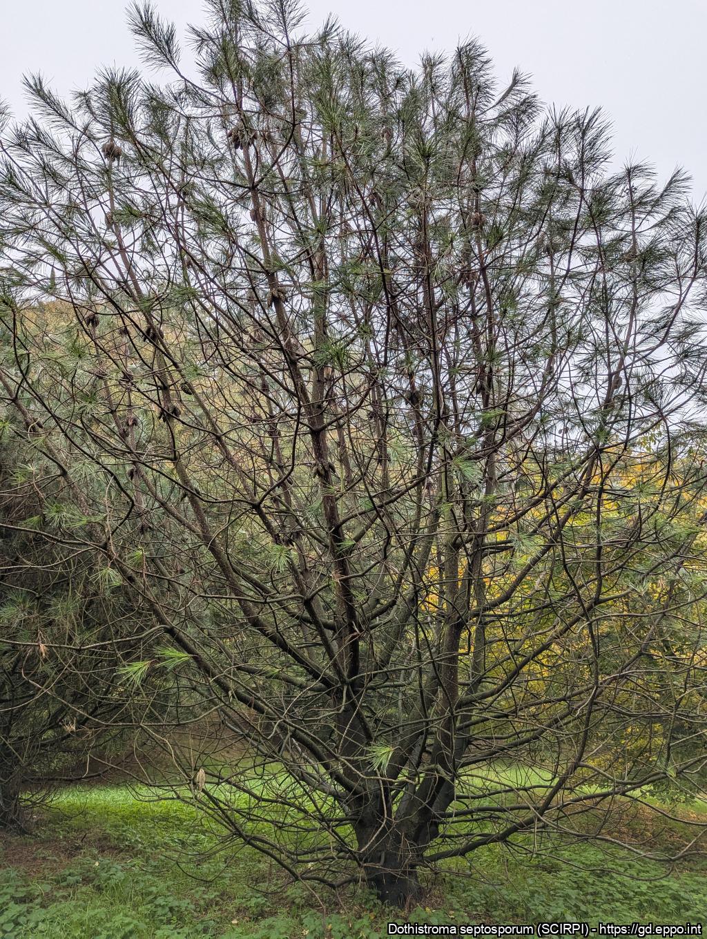
'Lion tail' symptoms on Pinus attenuata caused by Dothistroma sceptosporum (Yorkshire Arboretum, GB, 2024).
Courtesy: Camille PICARD (EPPO)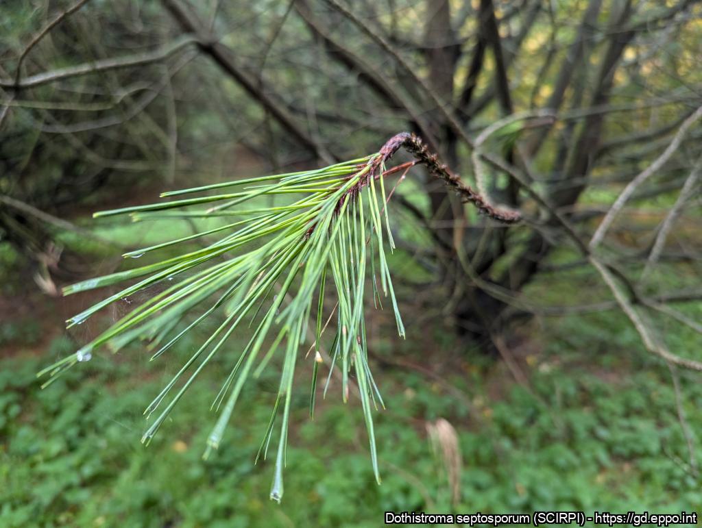
'Lion tail' symptoms on Pinus attenuata caused by Dothistroma sceptosporum (Yorkshire Arboretum, GB, 2024).
Courtesy: Camille PICARD (EPPO)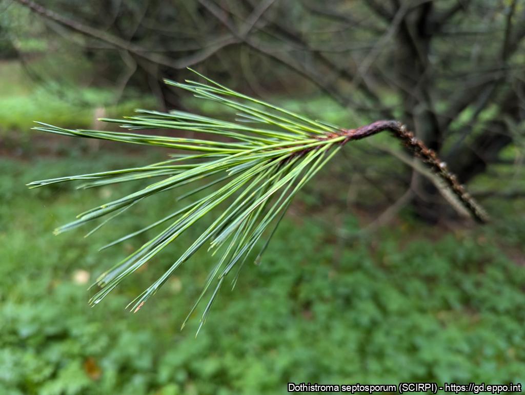
'Lion tail' symptoms on Pinus attenuata caused by Dothistroma sceptosporum (Yorkshire Arboretum, GB, 2024).
Courtesy: Camille PICARD (EPPO)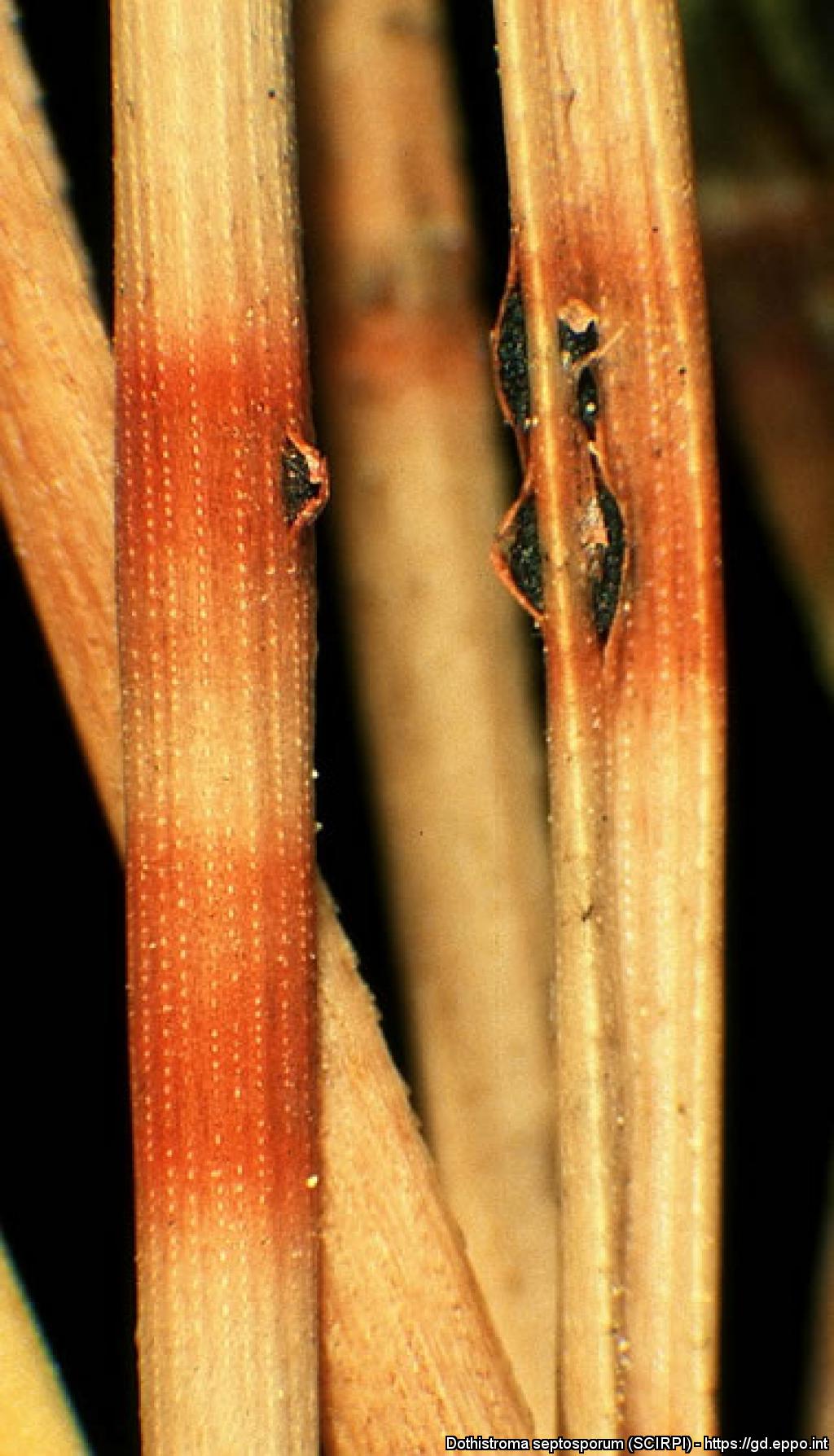
Symptoms on Pinus nigra: needles with brown necrotic tissue, showing typical red bands.
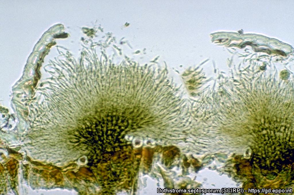
Dothistroma anamorph of M. pini; note prominent, aggregated acervuli and exposed conidiogenous tissues.
Courtesy: H.C. Evans, CABI Wallingford (GB).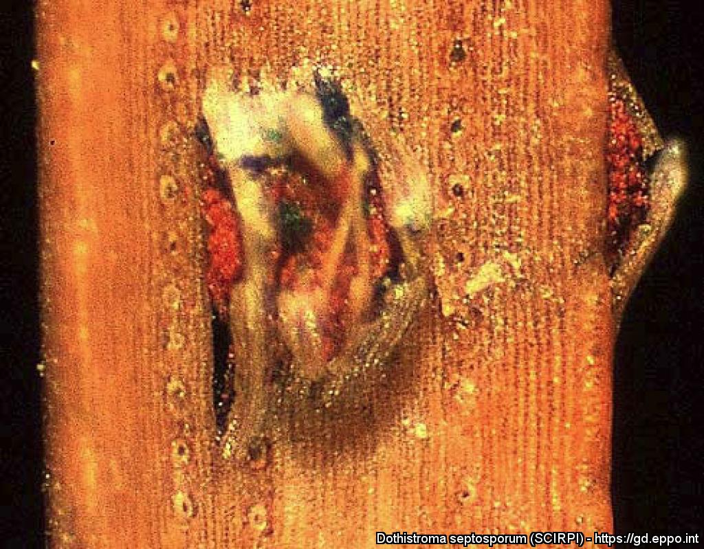
Fructifications of Mycosphaerella pini rupturing needle epidermis of Pinus mugo. Red pigments are visible (lens).
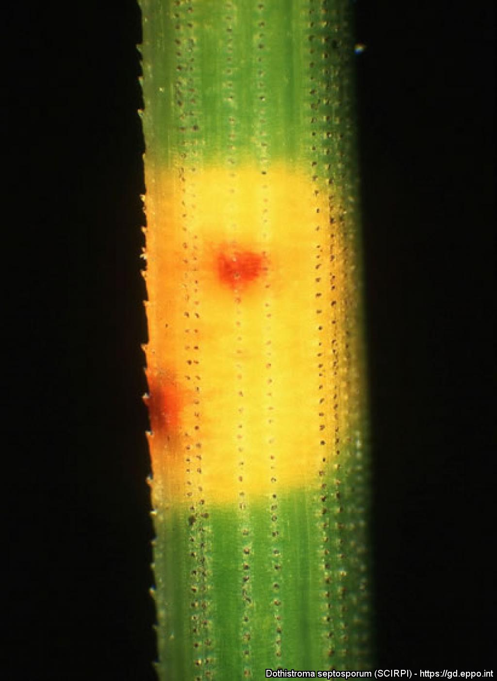
Yellow spots and necrotic bands on needles of Pinus mugo caused by Mycosphaerella pini (Dothistroma septospora).
Symptoms of Mycosphaerella pini: needles of Pinus mugo showing dead tips with green basis.
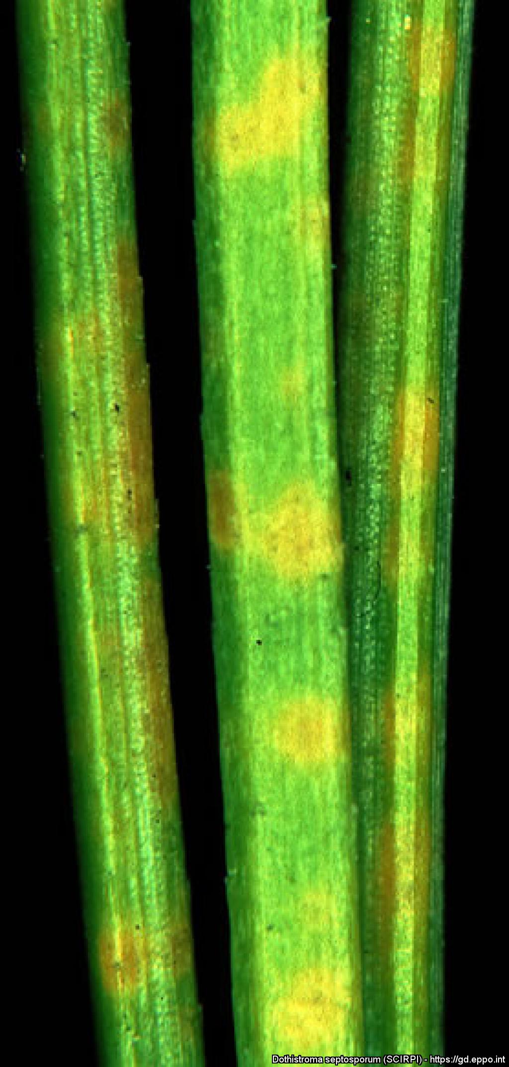
Yellow spots and necrotic bands on needles of Pinus cembra caused by Mycosphaerella pini (Dothistroma septospora).
Typical red bands on needles of Pinus nigra caused by Mycospaerella pini (Dothistroma septospora).
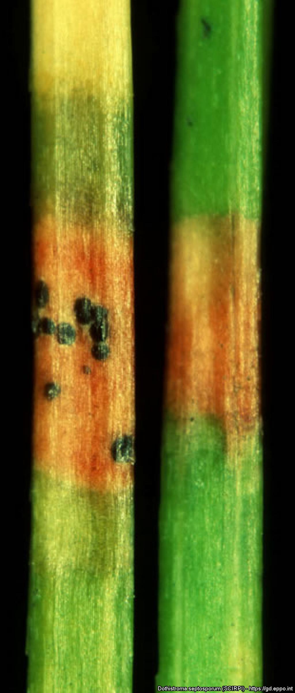
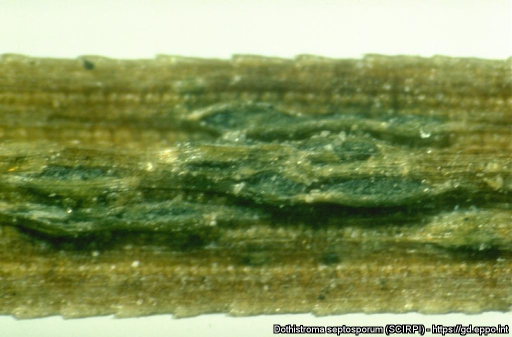
Symptoms of Mycophaerella pini: needles with brown necrotic tissue, showing black spots of stroma developing under the epidermis.
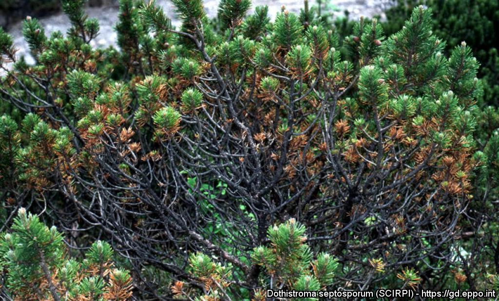
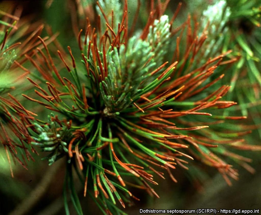
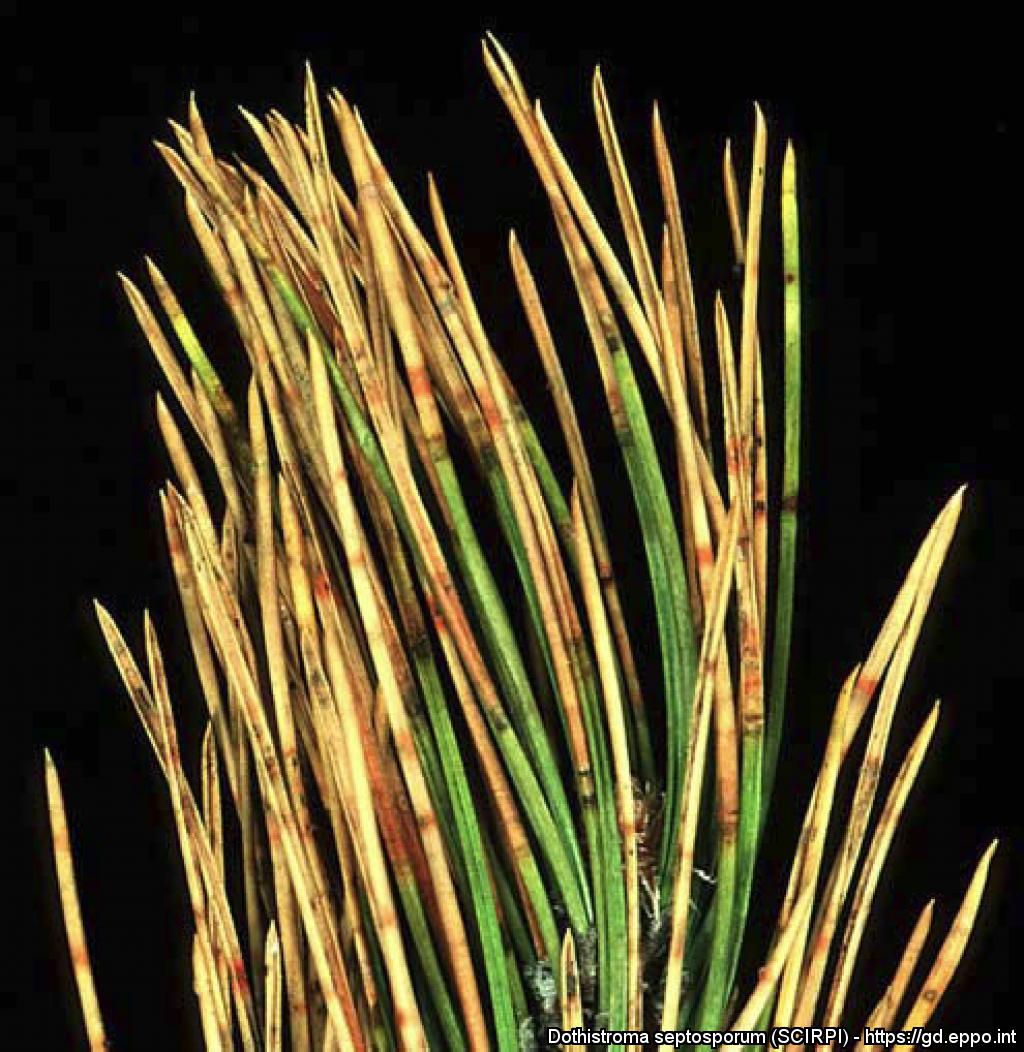
Attack of Mycosphaerella pini (Dothistroma septospora) on Pinus mugo over several years showing only last years needles (paintbrush-looking).

Symptoms of Mycophaerella pini: needles with brown necrotic tissue, showing fruit bodies breaking through the epidermis by longitudinal slits, raising a flap of epidermis.

Symptoms of Mycophaerella pini: needles with brown necrotic tissue, showing black spots of stroma developing under the epidermis.
Dothistroma (M. pini anamorph) acervulus (transverse section); note hyaline mass.
Courtesy: H.C. Evans, CABI Wallingford (GB).
M. pini (red band needle blight) on Pinus radiata, showing infection of both young and old foliage.
Courtesy: H.C. Evans, CABI Wallingford (GB).
M. pini on Pinus maximinoi; note aggregated, strongly erumpent ascostromata with needle reddening.
Courtesy: H.C. Evans, CABI Wallingford (GB).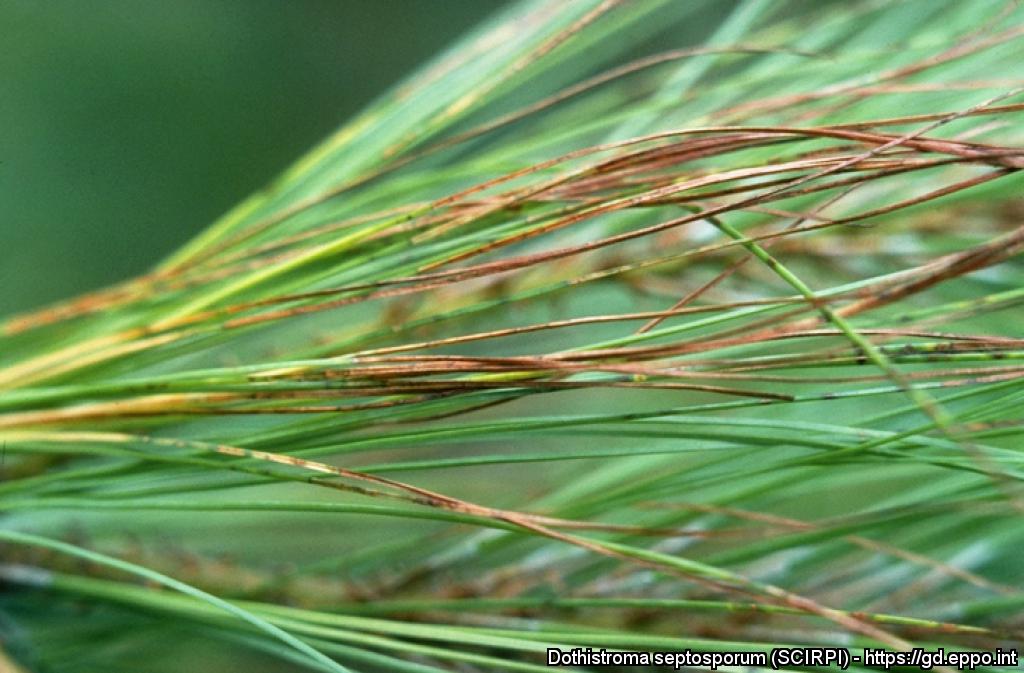
M. pini (red band needle blight) on Pinus maximinoi; note general reddening of needles.
Courtesy: H.C. Evans, CABI Wallingford (GB).