Lecanosticta acicola(SCIRAC)
Photos
For publication in journals, books or magazines, permission should be obtained from the original photographers with a copy to EPPO.
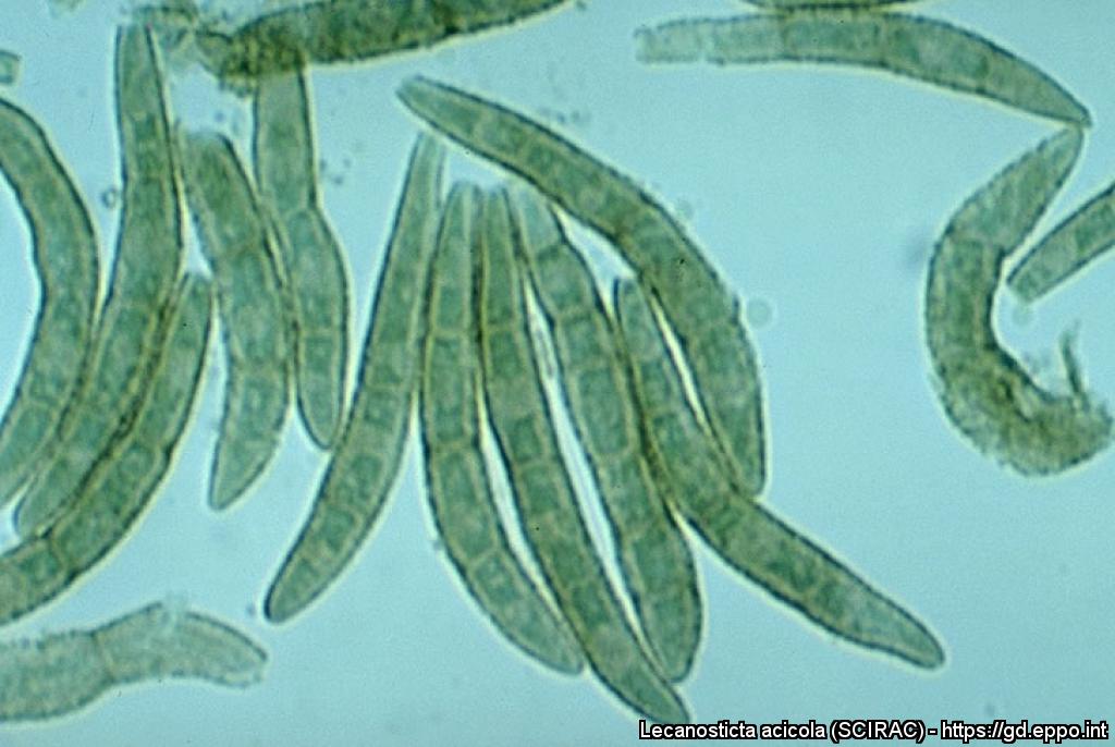
Lecanosticta conidia, to show variation in spore form with increasing altitude, on Pinus maximinoi (note decrease in size, septation, pigmentation and ornamentation with increasing altitude.
Courtesy: H.C. Evans, CABI, Wallingford (GB).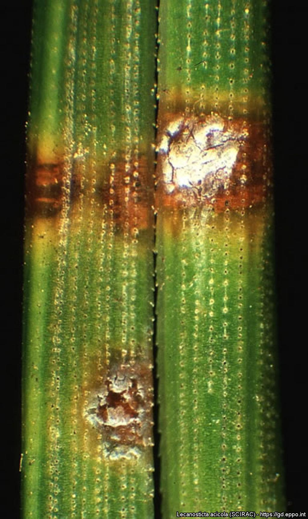
Brown spots and necrotic bands on needles (with resin-soaked spots) of Pinus mugo
Lecanosticta conidia, to show variation in spore form with increasing altitude, on Pinus maximinoi (note decrease in size, septation, pigmentation and ornamentation with increasing altitude.
Courtesy: H.C. Evans, CABI, Wallingford (GB).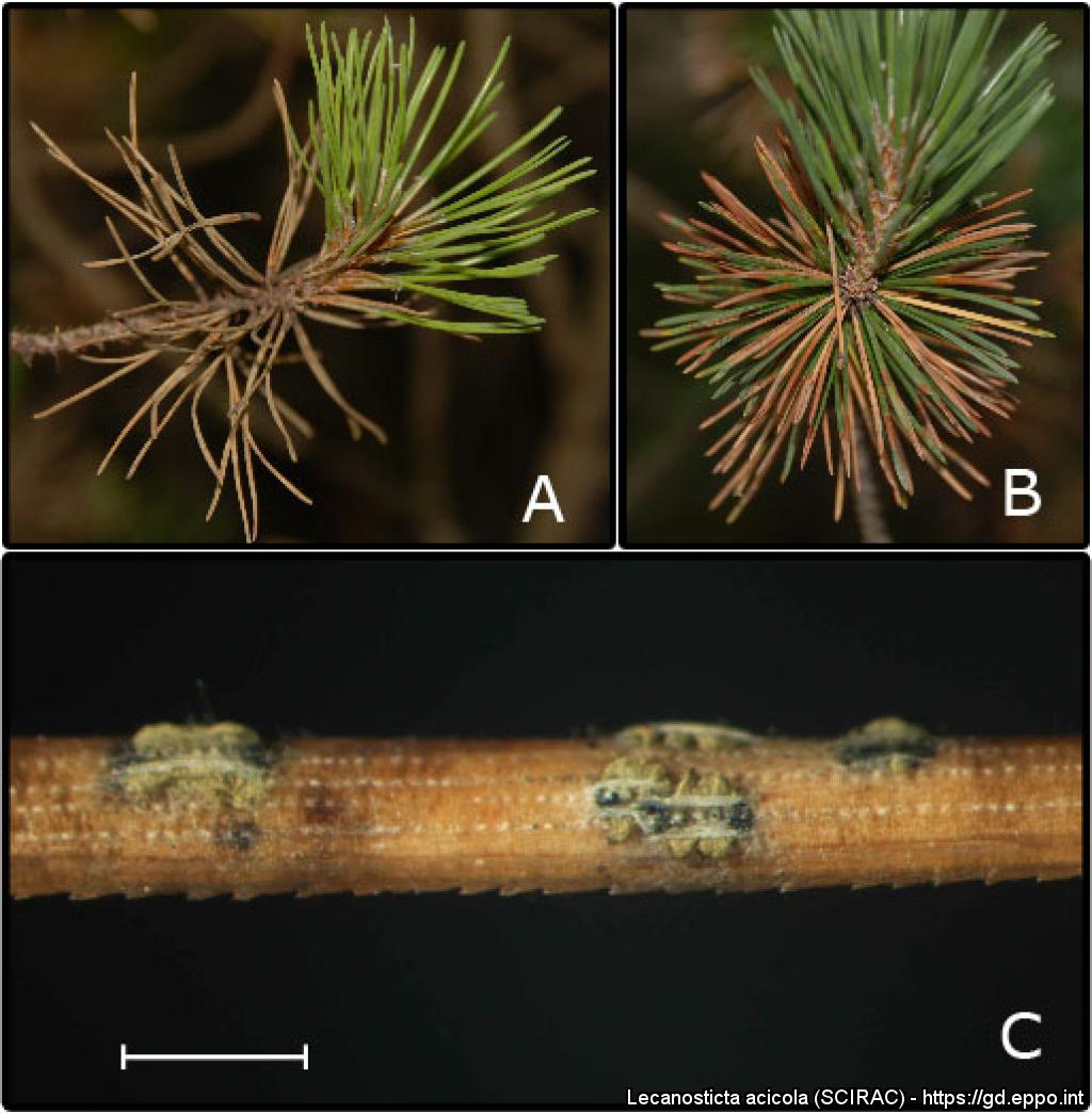
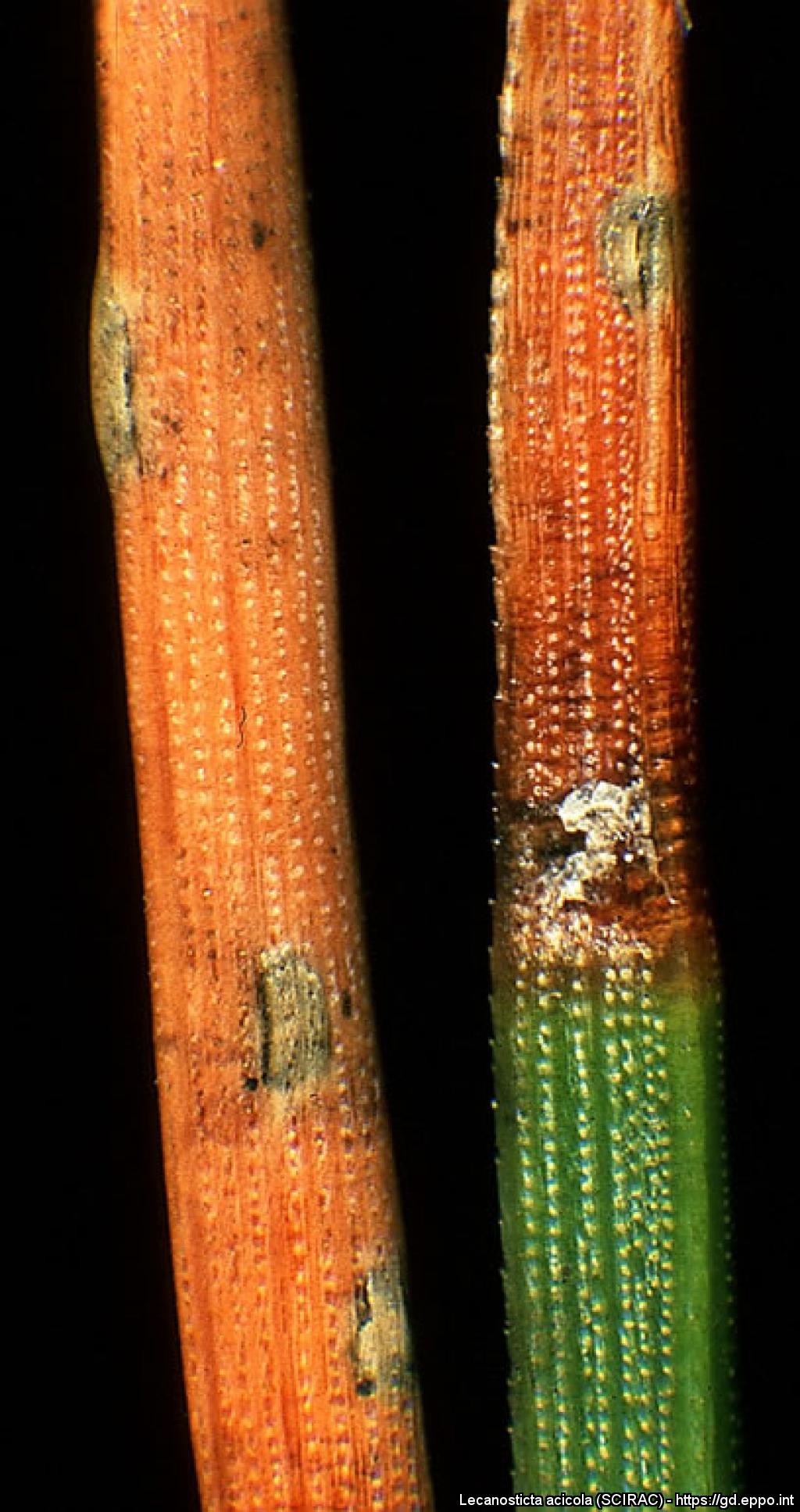
Brown to grey needles on Scotch pine (A) and stone pine (B) infected with brown-spot needle blight; conidial masses are protruding from both sides of the conidiomata under damp conditions (C) (bar 1 mm).
Courtesy: D Jurc, Forestry Institute, Ljubljana (SI).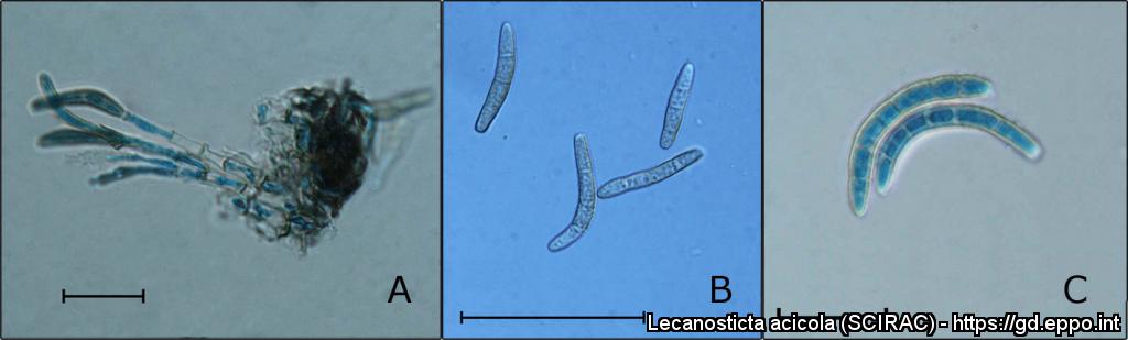
A. Stromatic hyphae, conidiophores, conidiogenous cells and developing conidia in lactophenol-cotton blue (bar 20 µm) - B. conidia in water showing light brown colouration and verrucose structure (bar 50 µm) - C. in lactophenol-cotton blue showing septa and a thickened wall ( bar 20 µm).
Courtesy: D Jurc, Forestry Institute, Ljubljana (SI).
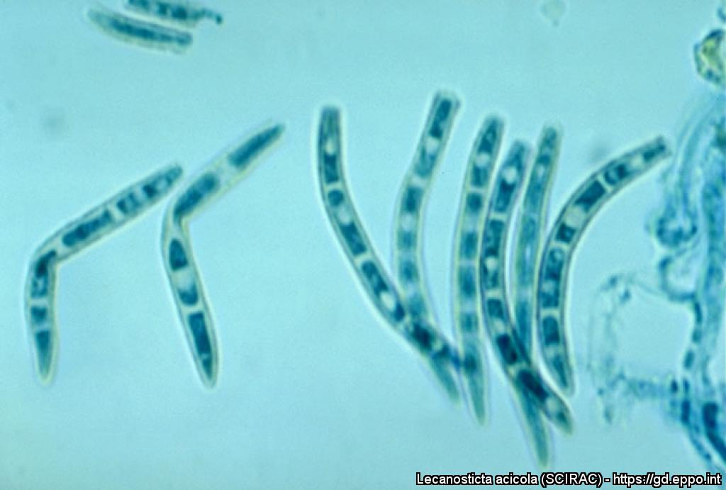
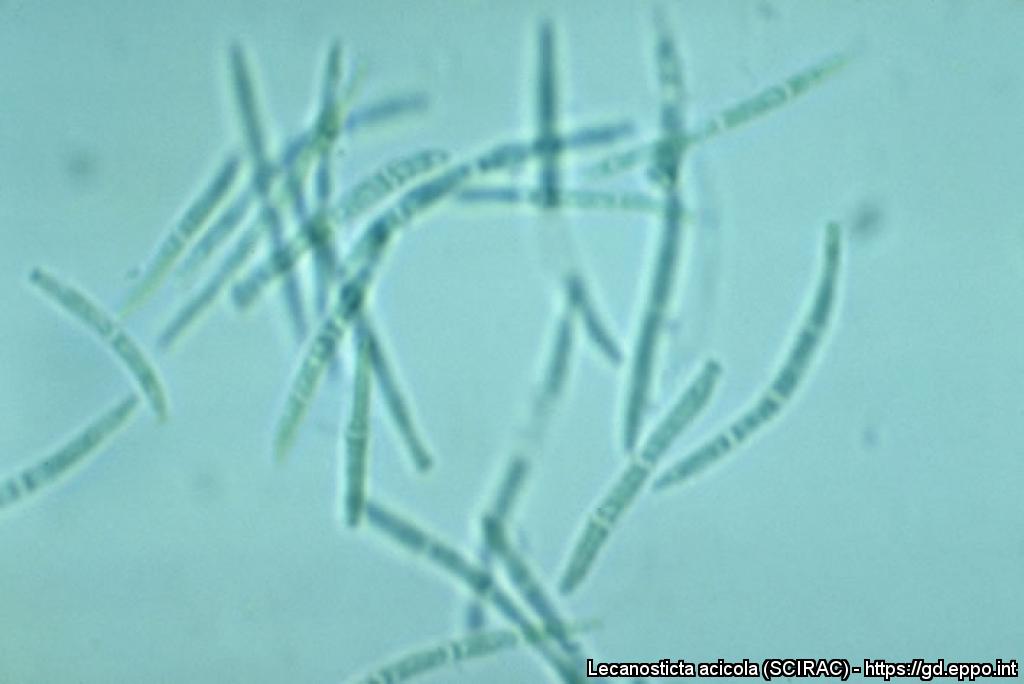
Conidia of Lecanosticta acicola.

Attack of Mycosphaerella dearnessii (Lecanosticta acicola) on Pinus mugo.
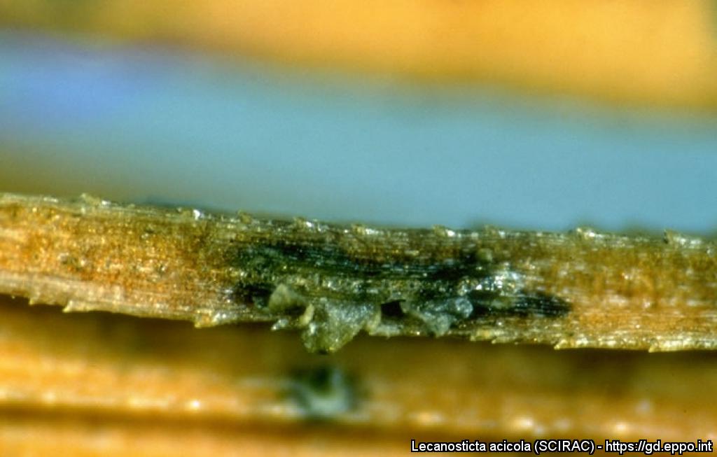
Lecanosticta anamorph of M. dearnessii; note solitary, discrete acervulus with green conidial cirrhus exuding onto needle.
Courtesy: H.C. Evans, CABI, Wallingford (GB).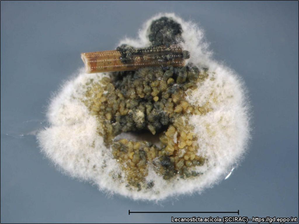
Mycelium from an infected needle with olive green conidial masses after 15 days from isolation on malt extract agar (bar 5 mm).
Courtesy: D Jurc, Forestry Institute, Ljubljana (SI).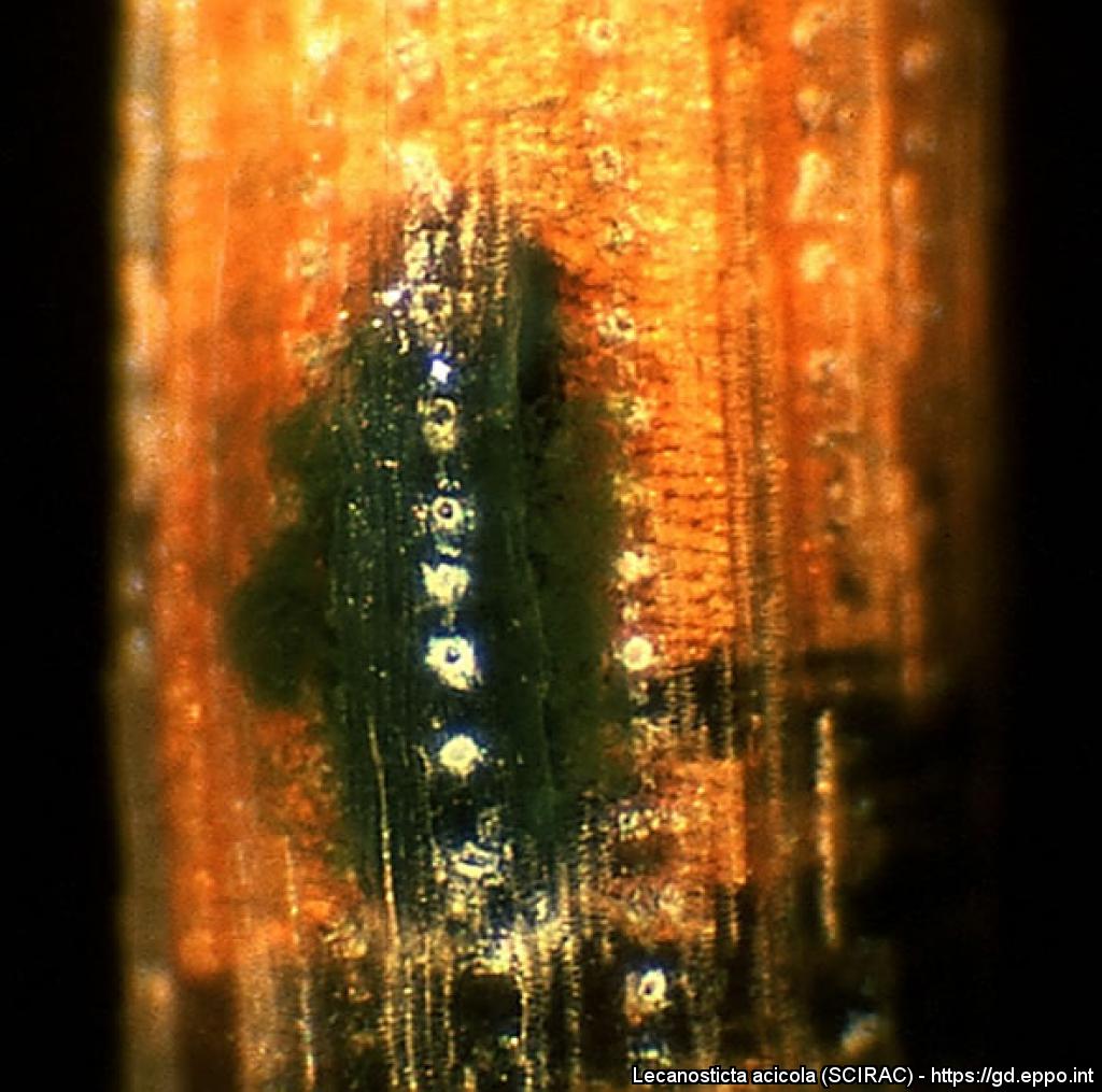
Slimy-dark green spore mass of Lecanosticta acicola produced from conidiomata under moist condition.
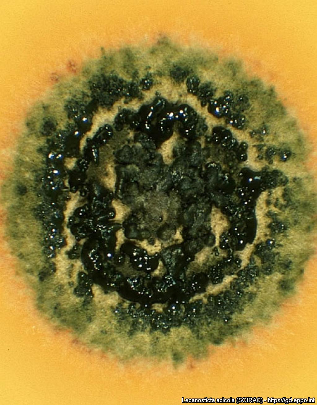
Sporulating culture of Lecanosticta acicola on malt extract agar.
Fructifications of Lecanosticta acicola rupturing needle epidermis of Pinus mugo.

Ascospores
Courtesy: H.C. Evans, CABI, Wallingford (GB).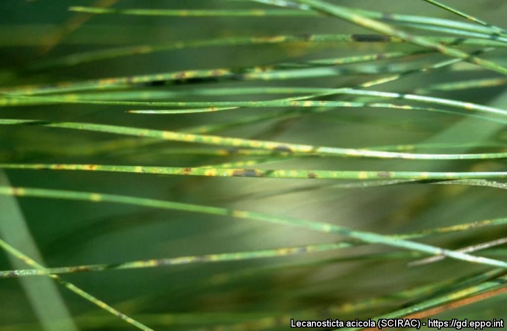
Close-up of brown spot symptoms on Pinus oocarpa.
Courtesy: H.C. Evans, CABI, Wallingford (GB).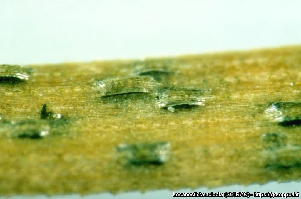
Lecanosticta acicola on Pinus oocarpa; note linearly arranged, solitary erumpent ascostromata.
Courtesy: H.C. Evans, CABI, Wallingford (GB).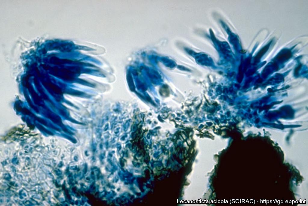
Asci
Courtesy: H.C. Evans, CABI, Wallingford (GB).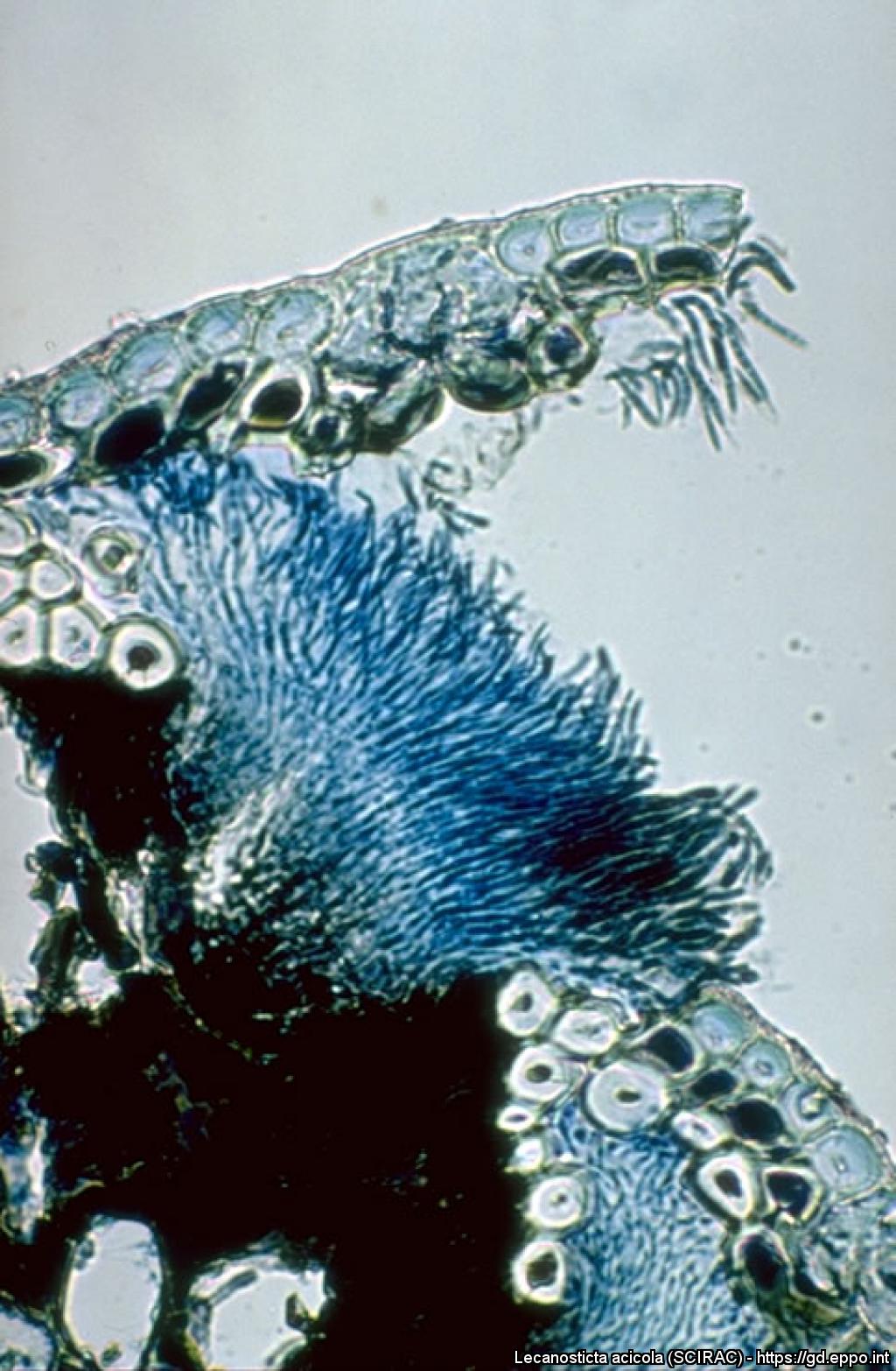
Acervulus (transverse section); note dark (green) spore mass.
Courtesy: H.C. Evans, CABI, Wallingford (GB).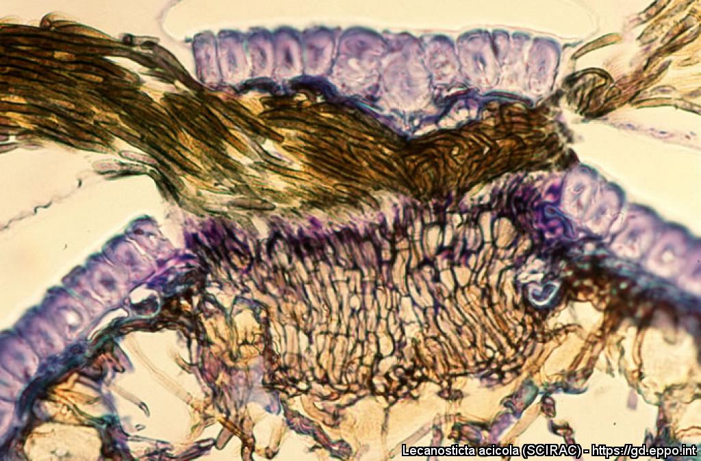
Cross section through a conidioma of Lecanosticta acicola (staining: thionine).
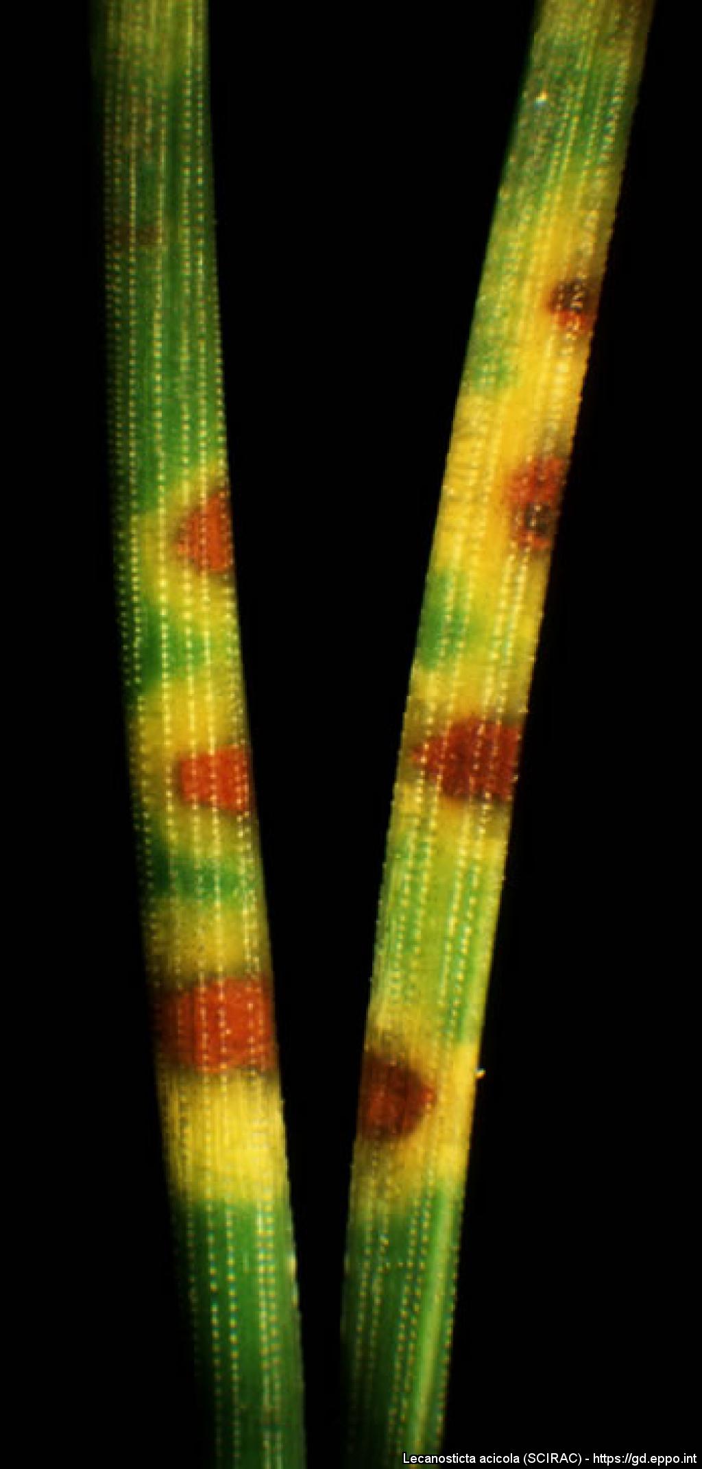
Brown spots and necrotic bands on needles of Pinus mugo
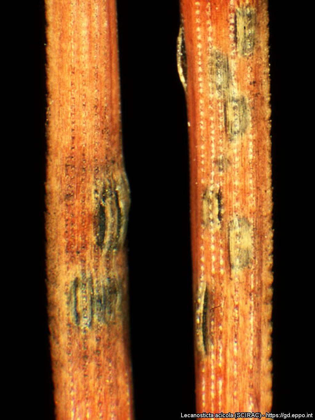
Fructifications of Lecanosticta acicola rupturing needle epidermis of Pinus mugo.
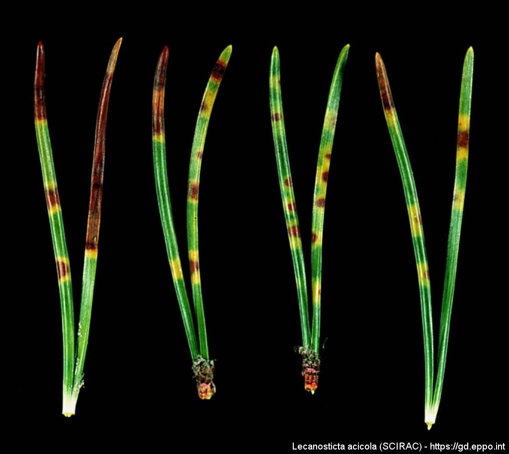
Brown spots and necrotic bands on needles of Pinus mugo
Dothistroma, conidia, from Pinus radiata; note hyaline, smooth, filiform, 1-3 septate spores.
Courtesy: H.C. Evans, CABI, Wallingford (GB).
Acervuli on Pinus sylvestris needles.
Courtesy: D.D. Skilling (US).
Infection on a Pinus sylvestris seedling.
Courtesy: A.G. Kais (US).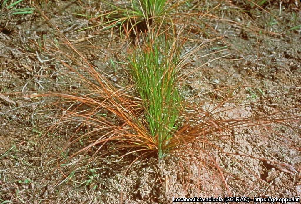
Infection on a Pinus palustris seedling.
Courtesy: A.G. Kais (US).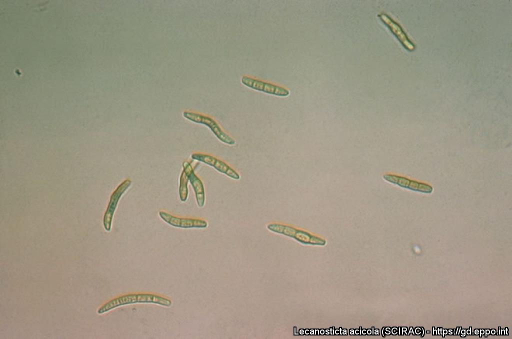
Conidia
Courtesy: A.G. Kais (US).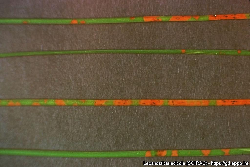
Close-up of an infection on Pinus sylvestris needles.
Courtesy: A.G. Kais (US).
Bbrown spot needle blight on second year foliage of Pinus maximinoi.
Courtesy: H.C. Evans, CABI, Wallingford (GB).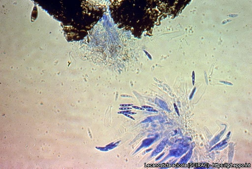
Perithecium, asci and ascospores
Courtesy: A.G. Kais (US).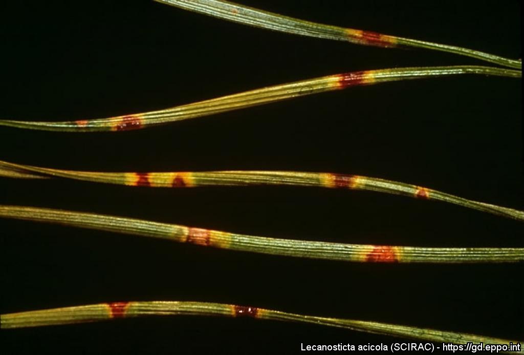
Typical lesions on Pinus sylvestris needles.
Courtesy: D.D. Skilling (US).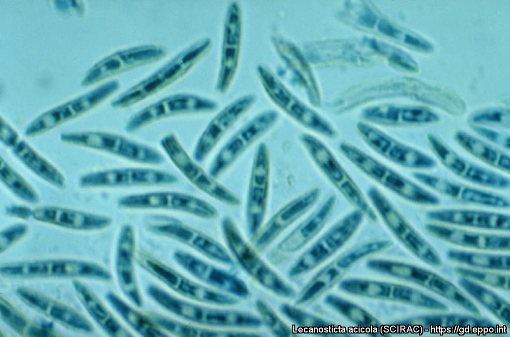
Conidia, to show variation in spore form with increasing altitude, on Pinus maximinoi (note decrease in size, septation, pigmentation and ornamentation with increasing altitude.
Courtesy: H.C. Evans, CABI, Wallingford (GB).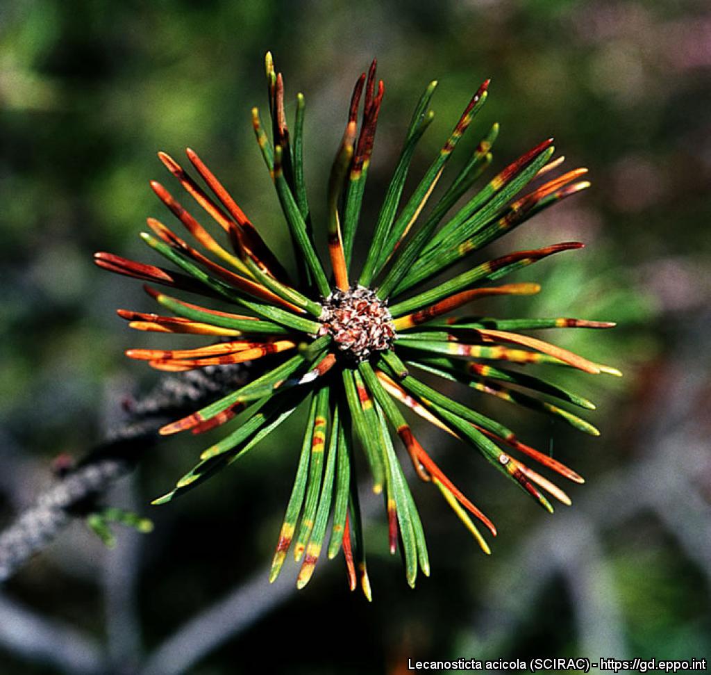
Symptoms on Pinus mugo.

Spots of black stroma of Lecanosticta acicola developing under the needle epidermis (Pinus mugo).
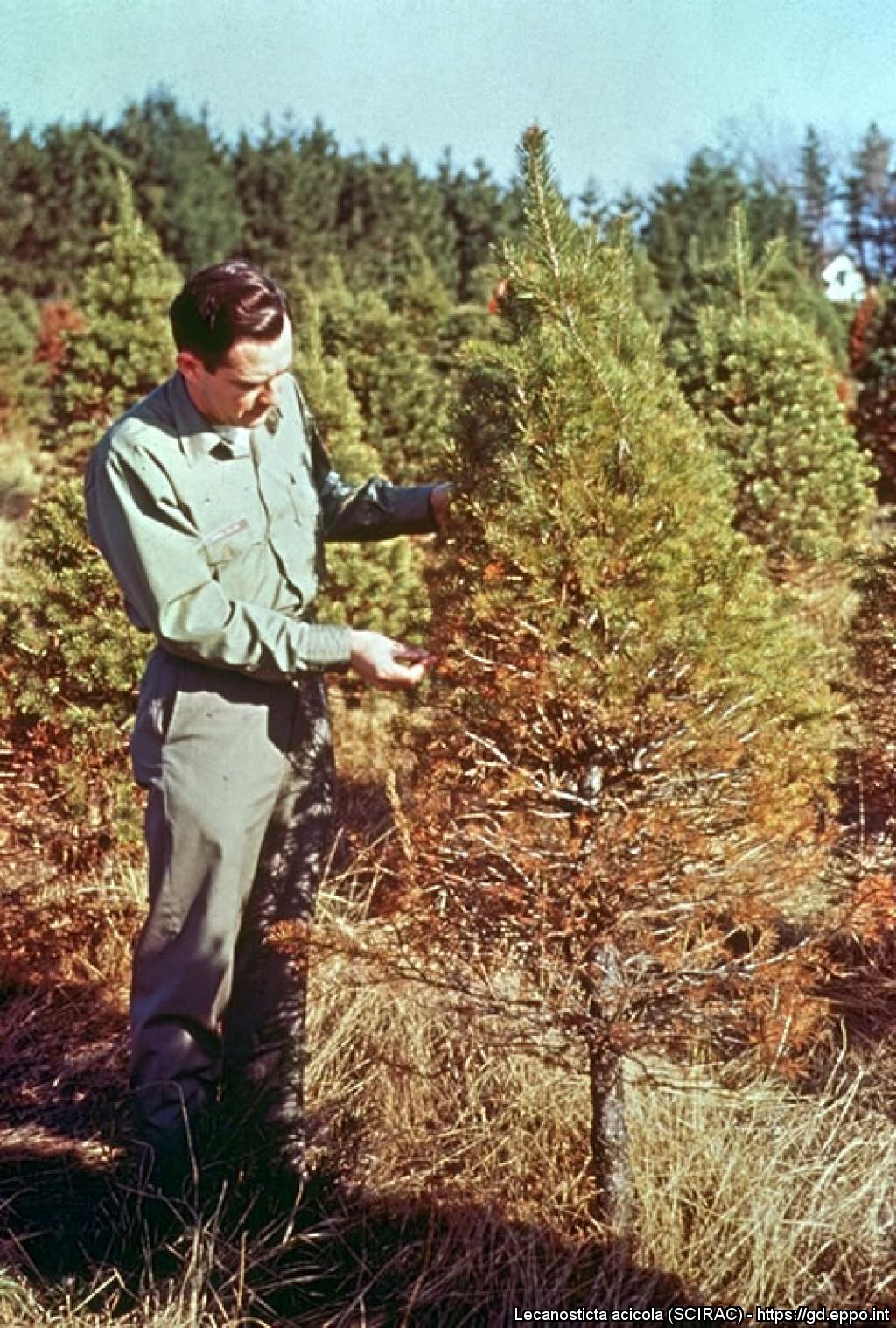
Symptoms on Pinus sylvestris; infection is more severe on the lower portion of the tree, especially on the north side where moisture conditions are most conducive to disease development.
Courtesy: D.D. Skilling (US).