Pseudomonas syringae pv. actinidiae(PSDMAK)
Photos
For publication in journals, books or magazines, permission should be obtained from the original photographers with a copy to EPPO.
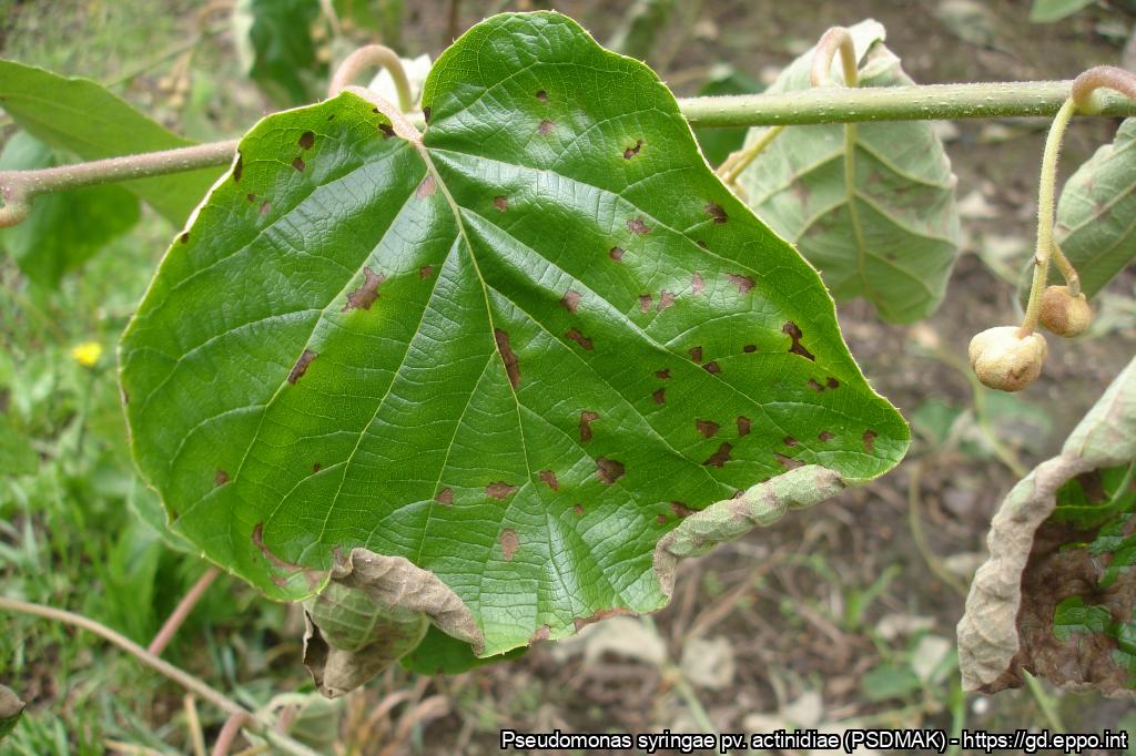
Symptoms on leaves
Courtesy: Carlos Coutinho - Entre Douro e Minho Plant Health Warning Station (Portugal)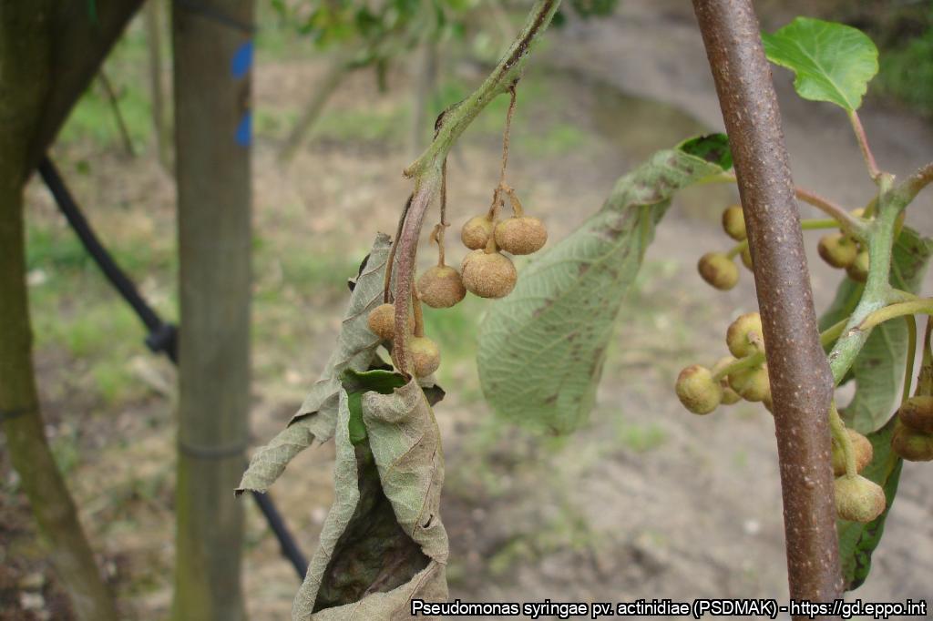
Dead flower buds
Courtesy: Carlos Coutinho - Entre Douro e Minho Plant Health Warning Station (Portugal)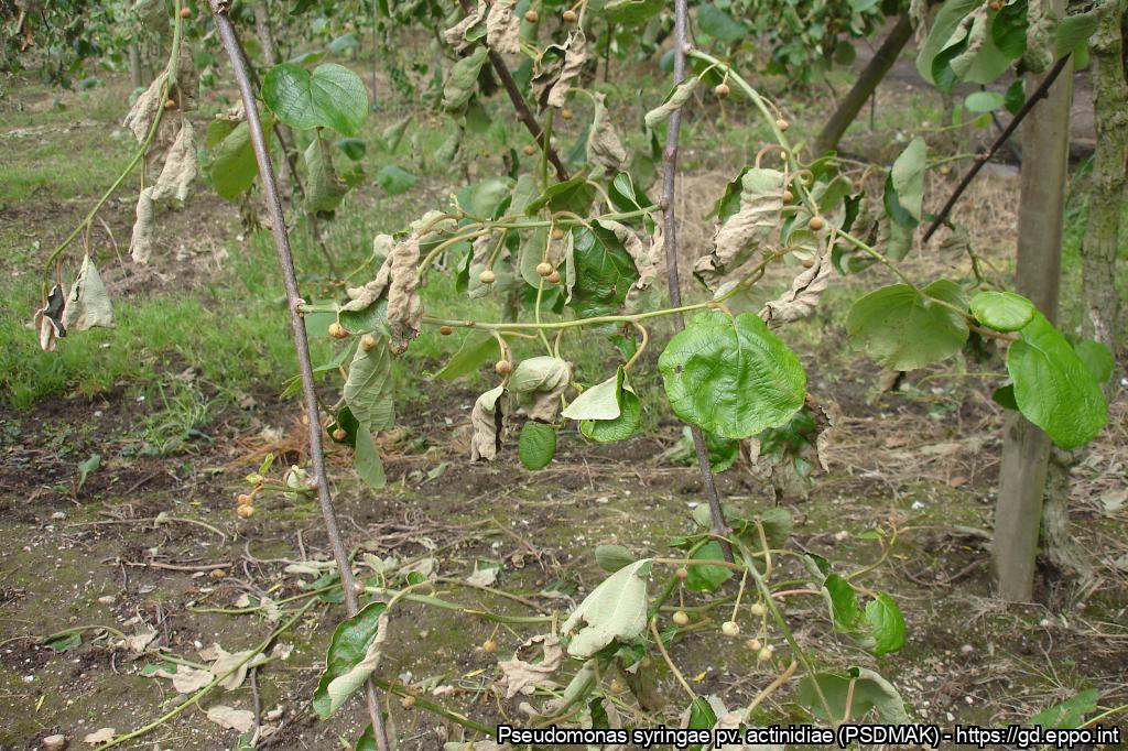
Affected leaves, branches and flower buds
Courtesy: Carlos Coutinho - Entre Douro e Minho Plant Health Warning Station (Portugal)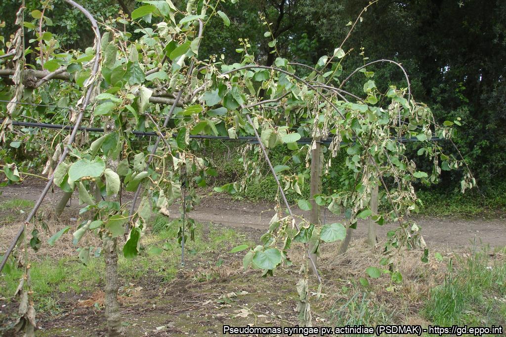
Plant very affected
Courtesy: Carlos Coutinho - Entre Douro e Minho Plant Health Warning Station (Portugal)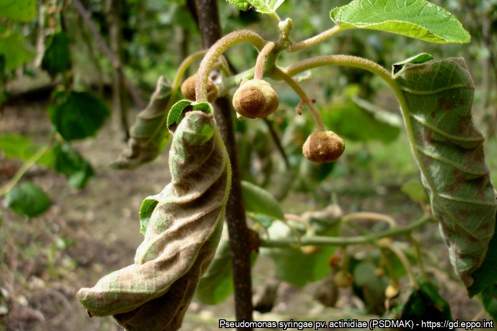
Attacked leaves and flower buds
Courtesy: Carlos Coutinho - Entre Douro e Minho Plant Health Warning Station (Portugal)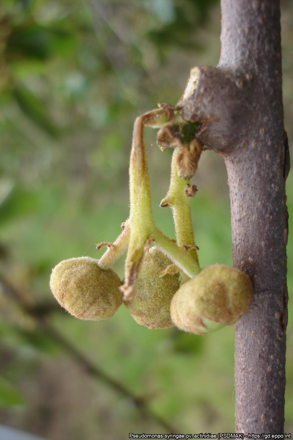
Symptoms on the flower bud stalk
Courtesy: Carlos Coutinho - Entre Douro e Minho Plant Health Warning Station (Portugal)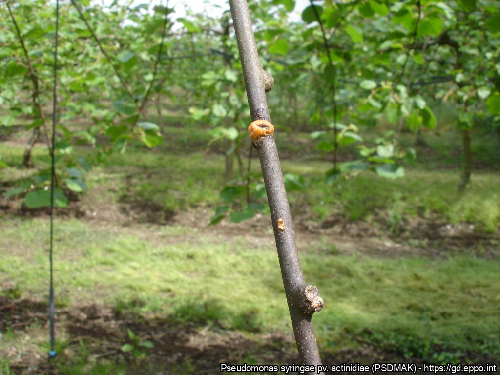
Bacterial exudate on the stick from the previous year
Courtesy: Carlos Coutinho - Entre Douro e Minho Plant Health Warning Station (Portugal)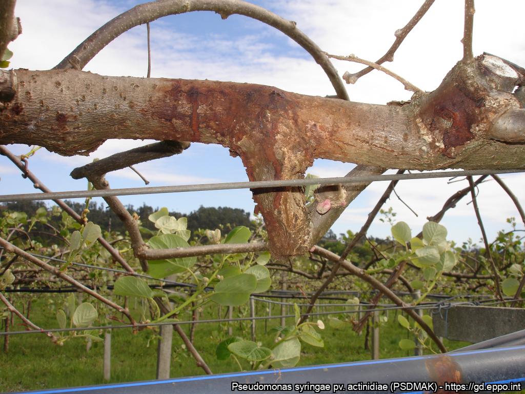
Bacterial exudate on the trunk at spring
Courtesy: Carlos Coutinho - Entre Douro e Minho Plant Health Warning Station (Portugal)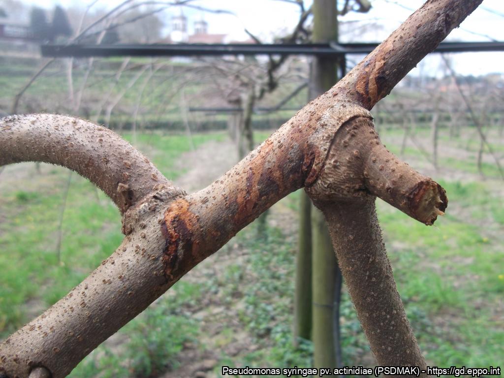
Beginning of the development of bacterial cankers on the secondary trunk
Courtesy: Carlos Coutinho - Entre Douro e Minho Plant Health Warning Station (Portugal)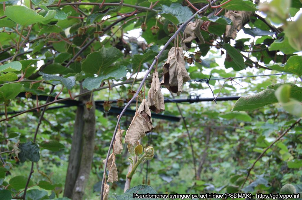
Dry, blackened twig
Courtesy: Carlos Coutinho - Entre Douro e Minho Plant Health Warning Station (Portugal)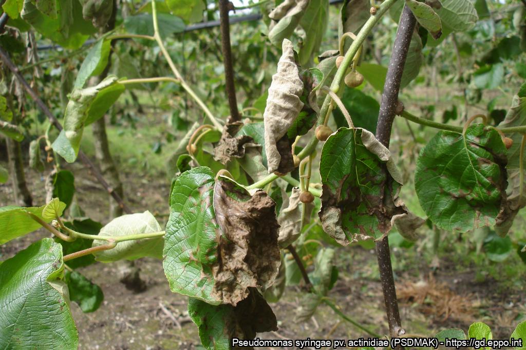
Symptoms on the leaves in a severely affected orchard
Courtesy: Carlos Coutinho - Entre Douro e Minho Plant Health Warning Station (Portugal)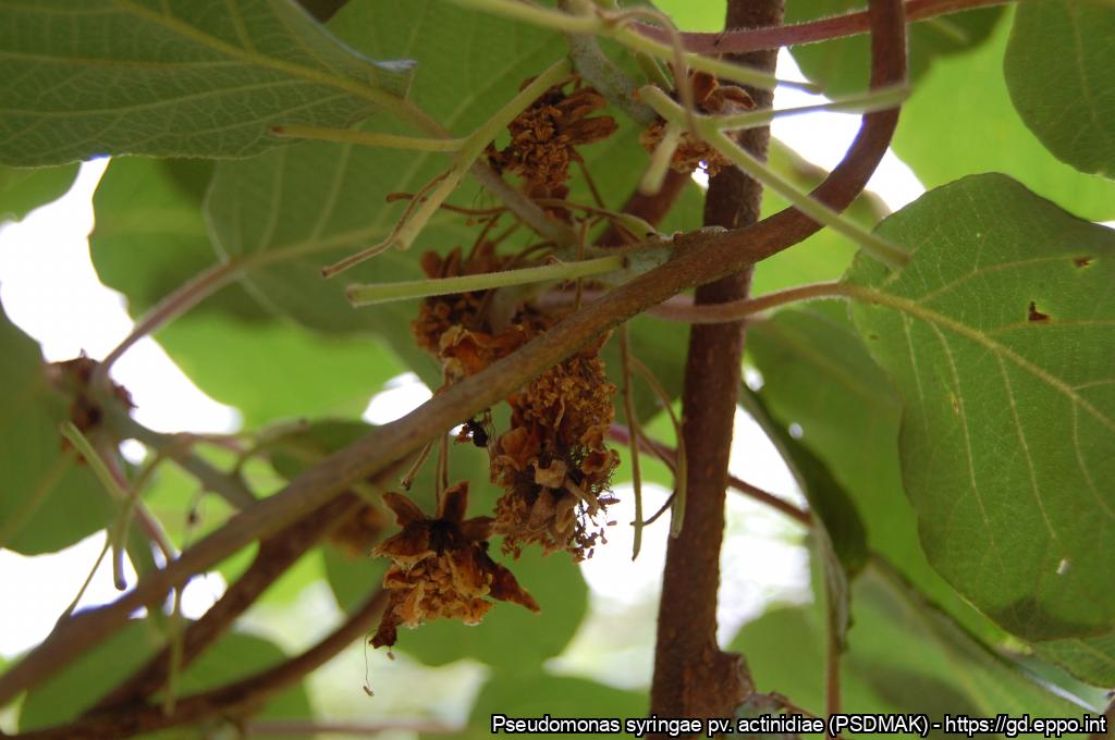
Aborted fruitlets as a result of PSA attack
Courtesy: Carlos Coutinho - Entre Douro e Minho Plant Health Warning Station (Portugal)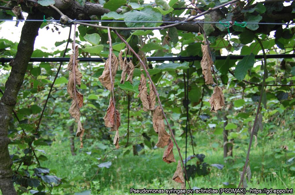
Branches of the year destroyed by PSA
Courtesy: Carlos Coutinho - Entre Douro e Minho Plant Health Warning Station (Portugal)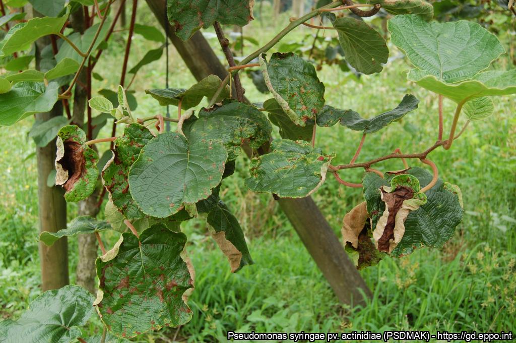
Symptoms on leaves at various stages of development
Courtesy: Carlos Coutinho - Entre Douro e Minho Plant Health Warning Station (Portugal)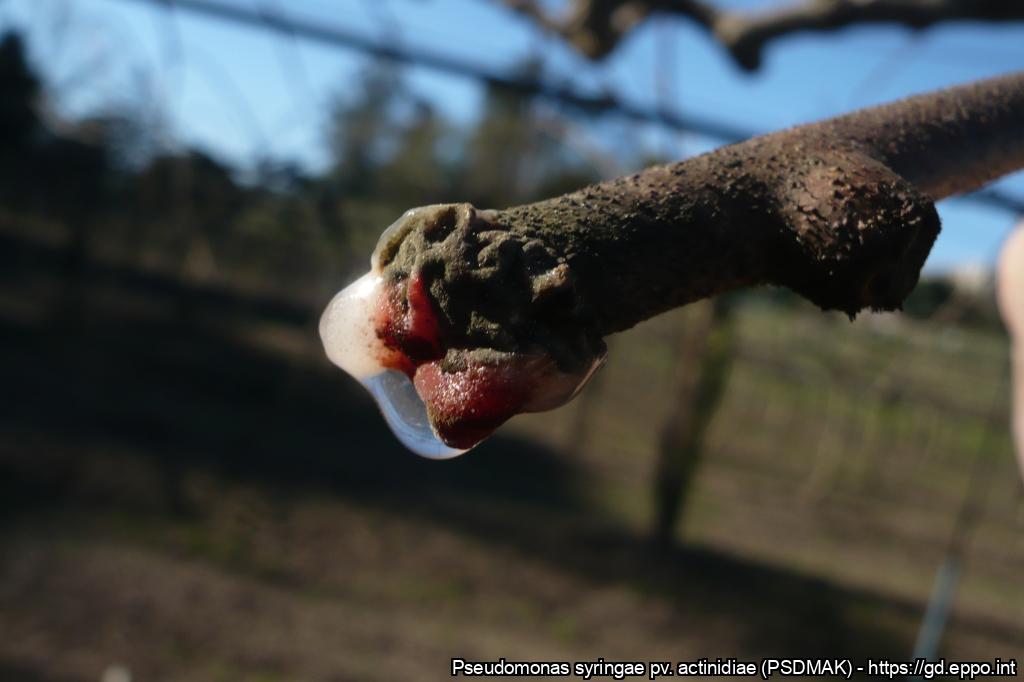
Bacterial exudate on the stick from the previous year
Courtesy: Carlos Coutinho - Entre Douro e Minho Plant Health Warning Station (Portugal)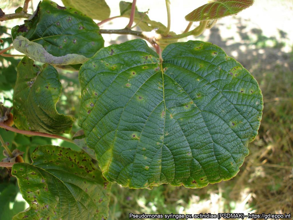
Symptoms on leaves
Courtesy: Carlos Coutinho - Entre Douro e Minho Plant Health Warning Station (Portugal)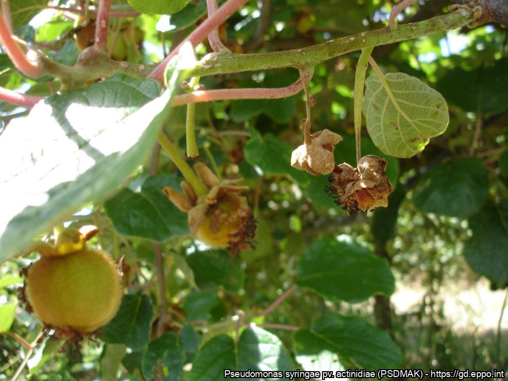
Aborted fruitlets as a result of PSA attack
Courtesy: Carlos Coutinho - Entre Douro e Minho Plant Health Warning Station (Portugal)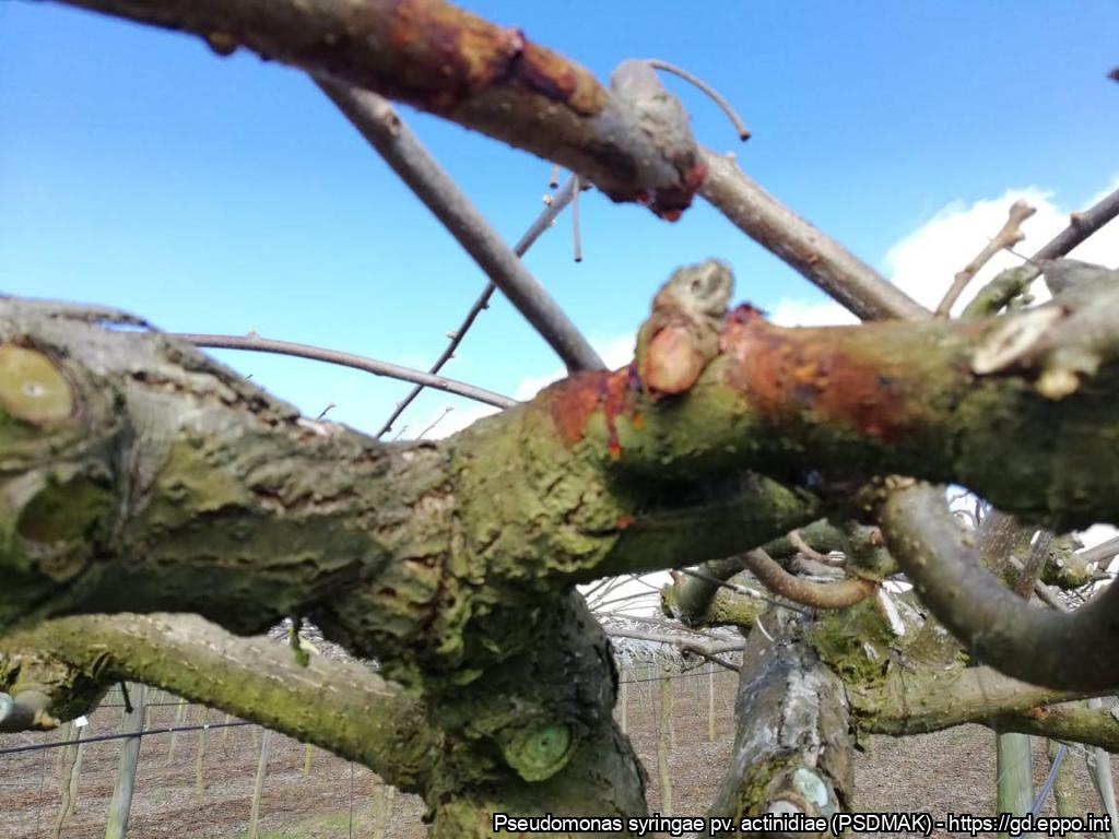
PSA Kiwis variety Hayward
Courtesy: Miguel Angel Fierro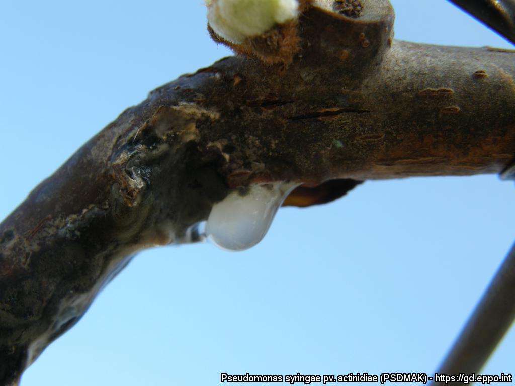
Bacterial ooze due to infection on kiwiplant
Courtesy: Riccardo Bugiani (Plant Protection Service - Emilia-Romagna Region - IT)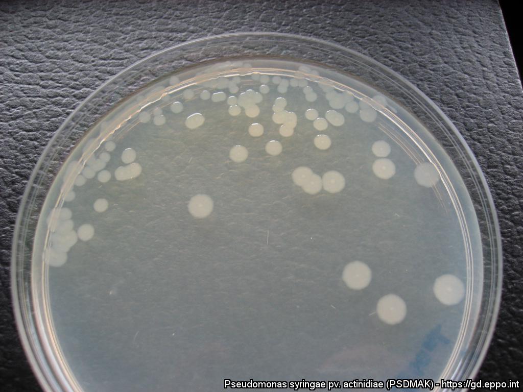
Morphology of colonies grown for 5 days on King’s medium B, supplemented with antibiotics
Courtesy: E. Stefani, Dept. of Life Sciences, Reggio Emilia (IT)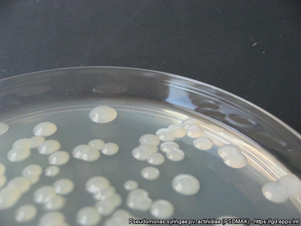
Morphology of colonies grown for 5 days on NSA medium, supplemented with antibiotics
Courtesy: E. Stefani, Dept. of Life Sciences, Reggio Emilia (IT)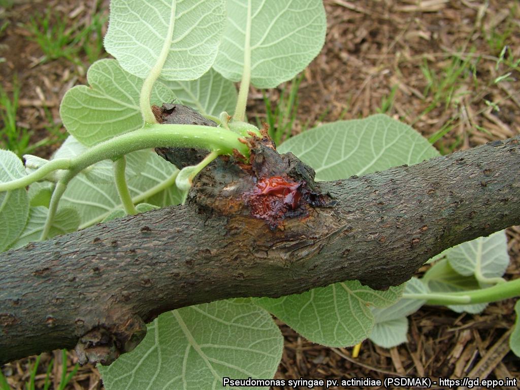
Bacterial exudate oozing from infected actinidia twig
Courtesy: Riccardo Bugiani (Plant Protection Service - Emilia-Romagna Region - IT)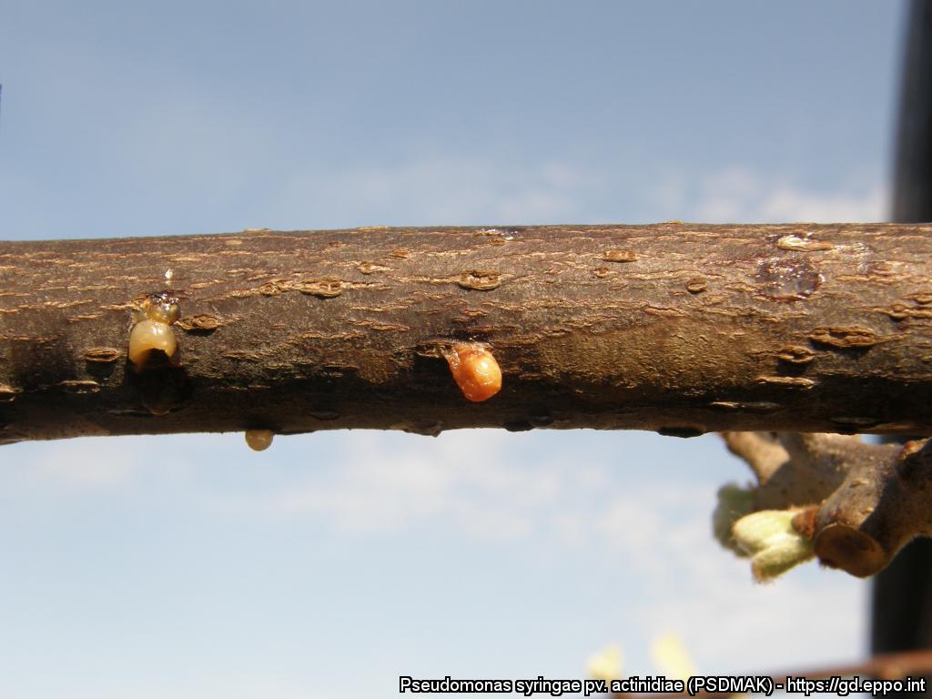
Bacterial exudate oozing from infected actinidia twigs
Courtesy: Riccardo Bugiani (Plant Protection Service - Emilia-Romagna Region - IT)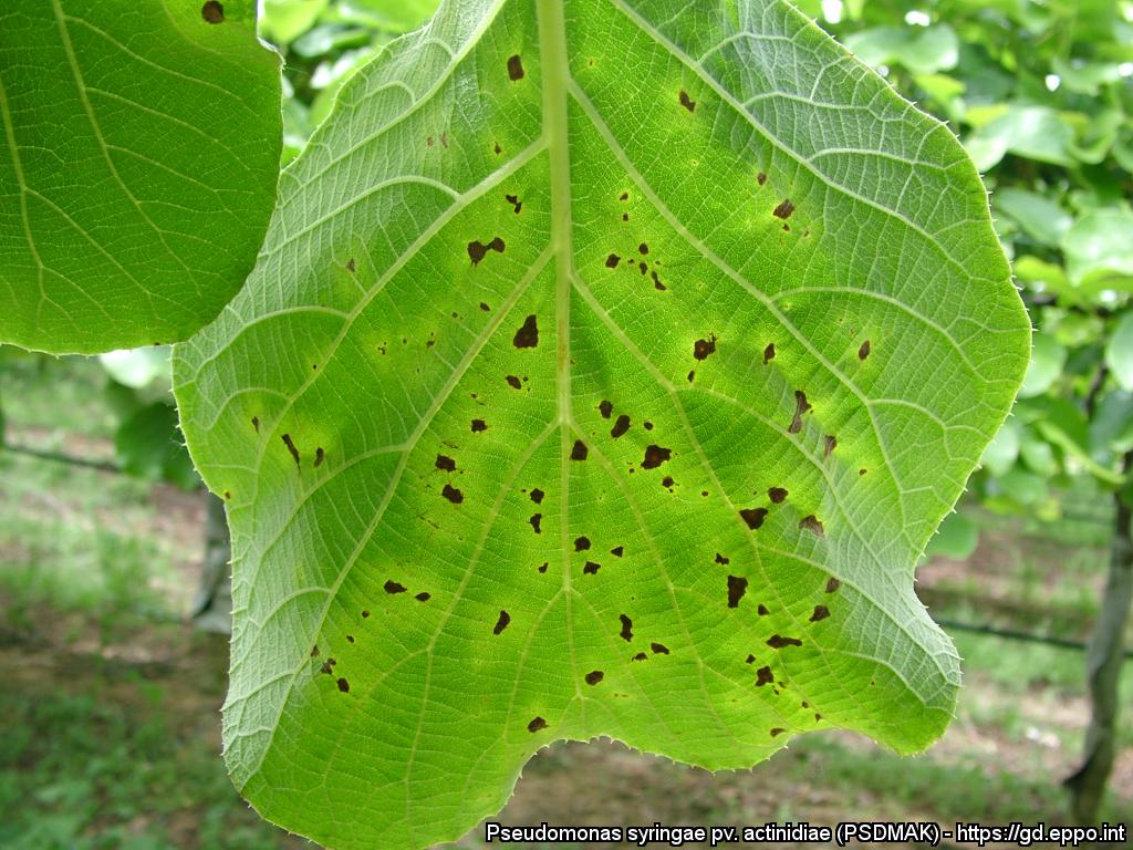
Brown spots surrounded by yellow haloes are visible in spring on kiwifruit leaves.
Courtesy: Plant Protection Service of Emilia-Romagna region (IT).
In spring, vine branches and trunks affected by Pseudomonas syringae pv. actinidiae show cankers which usually ooze red exudates.
Courtesy: Plant Protection Service of Emilia-Romagna region (IT).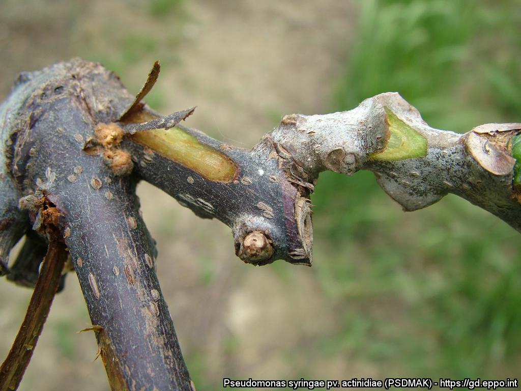
Browning of tissue under the cortex of vine branches (Actinidia chinensis).
Courtesy: Plant Protection Service of Emilia-Romagna region (IT).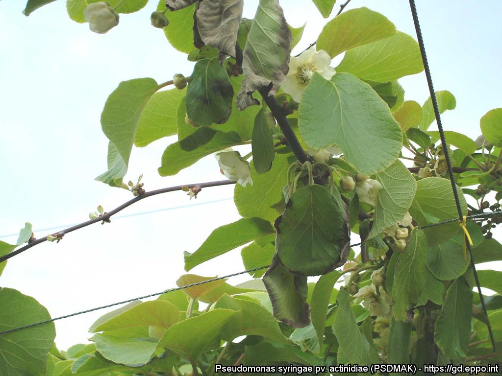
Colonization of vascular tissues by Pseudomonas syringae pv. actinidiae can result in foliar wilting at the beginning of the season (A. chinensis cv. Hort 16 A).
Courtesy: Plant Protection Service of Emilia-Romagna region (IT).