Heterodera elachista(HETDEL)
Photos
All photos included on this page can only be used for educational purposes.
For publication in journals, books or magazines, permission should be obtained from the original photographers with a copy to EPPO.
For publication in journals, books or magazines, permission should be obtained from the original photographers with a copy to EPPO.
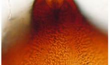
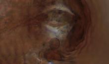
Terminal vulval cone. Scale bar = 50 µm.
Courtesy: Pablo Castillo - Institute for Sustainable Agriculture, CSIC, Cordoba (ES)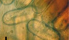
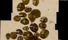
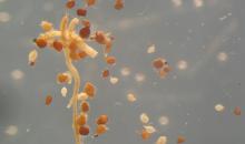
Detail of cyst contained embryos. Scale bar = 50 µm.
Courtesy: Pablo Castillo - Institute for Sustainable Agriculture, CSIC, Cordoba (ES)
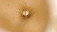
Fenestral cone features at different focus. Scale bar = 50 µm.
Courtesy: Pablo Castillo - Institute for Sustainable Agriculture, CSIC, Cordoba (ES)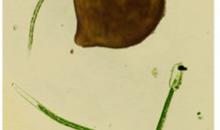
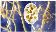
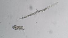
Juveniles and a mature cyst. Scale bar = 200 µm.
Courtesy: Pablo Castillo - Institute for Sustainable Agriculture, CSIC, Cordoba (ES)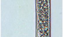
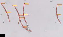
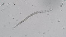
Entire body of second stage juvenile of Heteredera elachista. Scale bar = 50 µm.
Courtesy: Pablo Castillo - Institute for Sustainable Agriculture, CSIC, Cordoba (ES)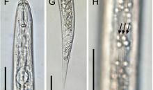
Anterior (F) and posterior (G) body portions, and detailed view of lateral field (H) of second stage juvenile. Scale bar = 20 µm.
Courtesy: Pablo Castillo - Institute for Sustainable Agriculture, CSIC, Cordoba (ES)
