Gymnosporangium sabinae(GYMNFU)
Photos
For publication in journals, books or magazines, permission should be obtained from the original photographers with a copy to EPPO.

Developing spermagonia on the upper surface of a pear leaf
Courtesy: Kostas Anastasopoulos (Department of Agriculture, I.H.U)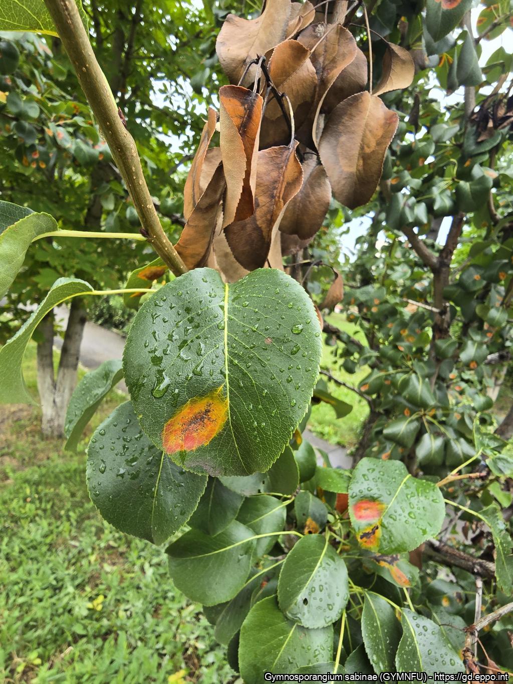
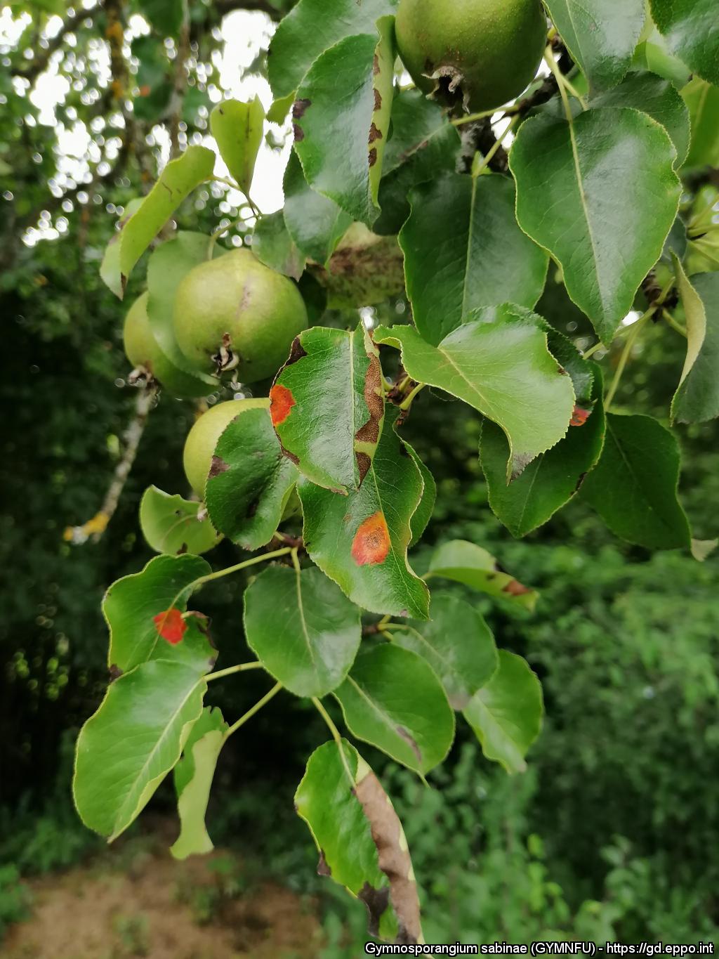
Pear rust: clear symptoms of the pathogen's presence. Russian Federation
Courtesy: Dr. Valentino De Rosa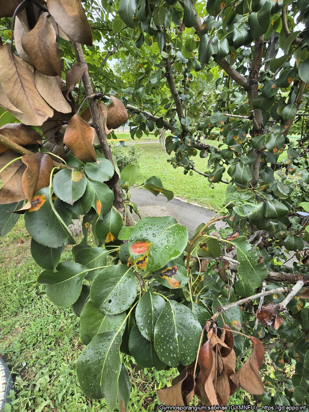
Pear rust: clear symptoms of the pathogen's presence. Russian Federation
Courtesy: Dr. Valentino De Rosa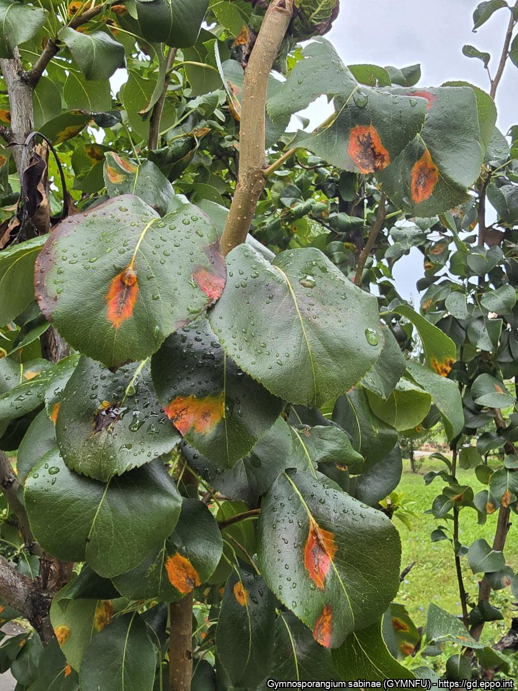
Pear rust: clear symptoms of the pathogen's presence. Russian Federation
Courtesy: Dr. Valentino De Rosa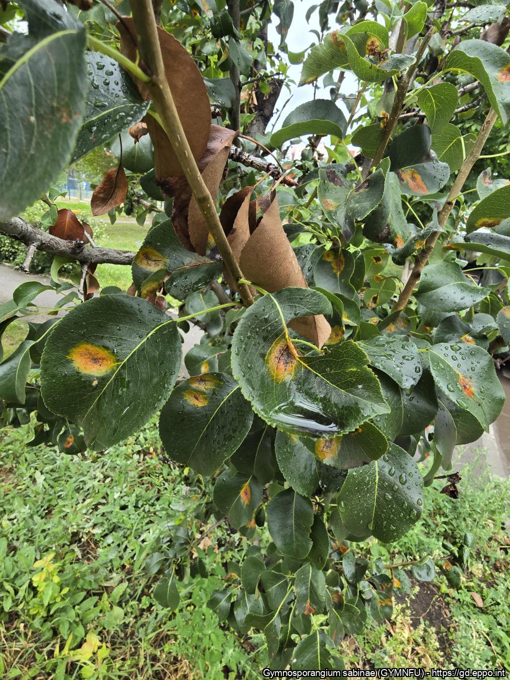
Pear rust: clear symptoms of the pathogen's presence. Russian Federation
Courtesy: Dr. Valentino De Rosa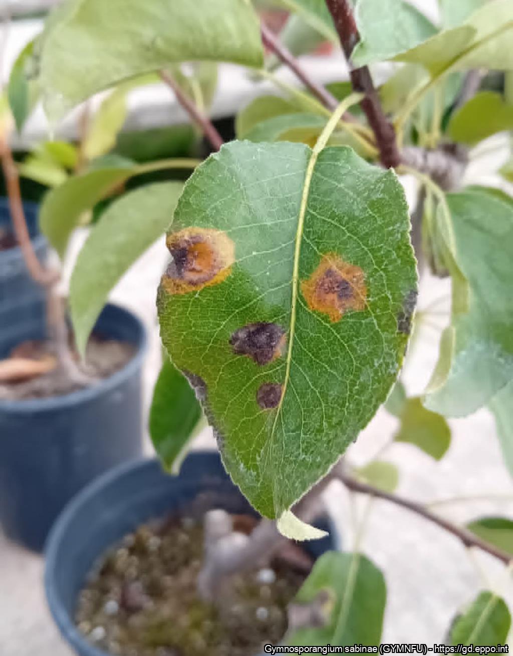
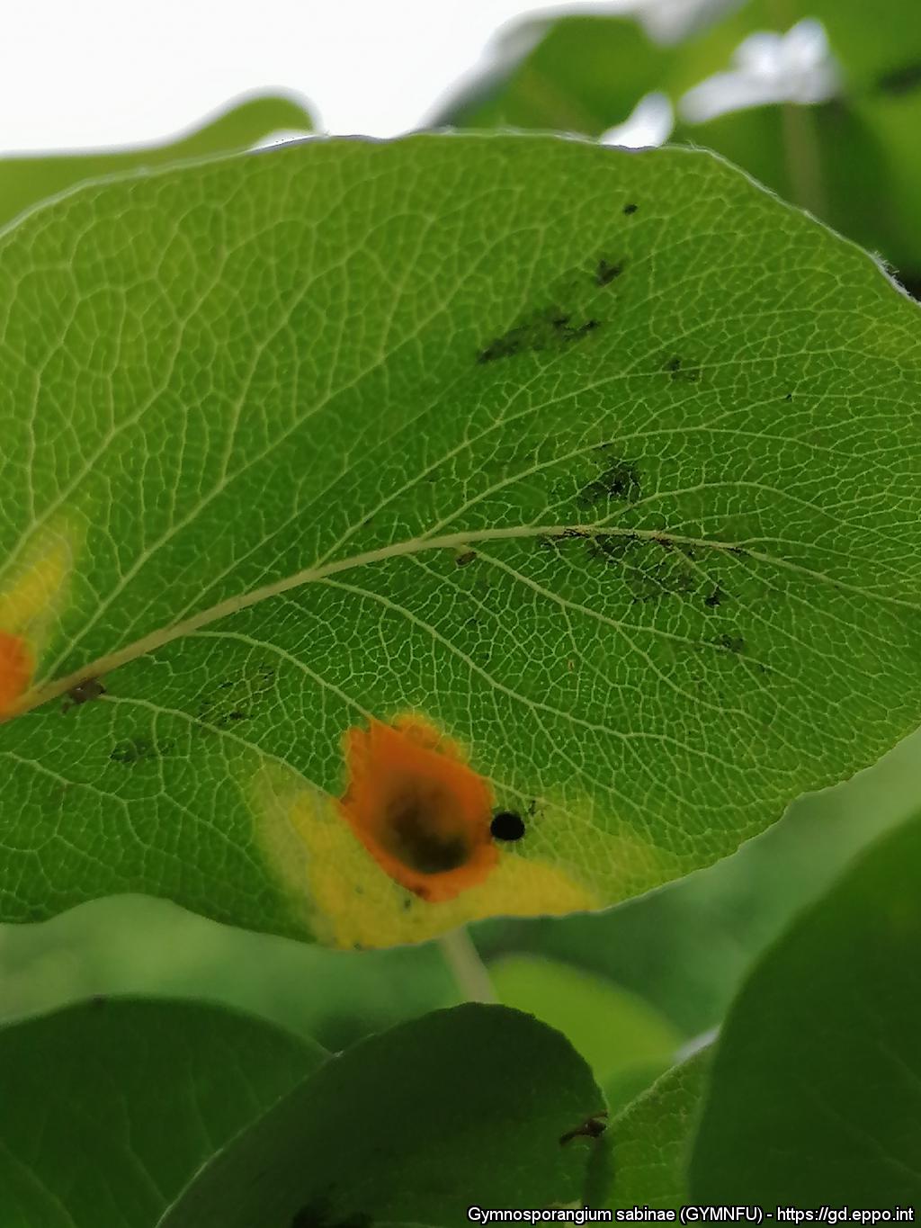
Later stage of the rust disease with large spots (developed spermagonia) on upper leaf surface
Courtesy: Kostas Anastasopoulos (Department of Agriculture, I.H.U)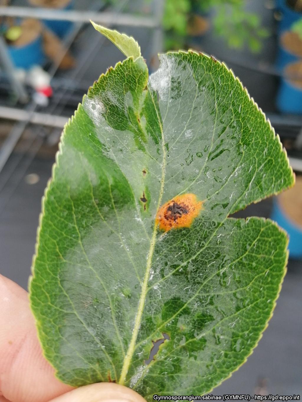
Developed aecia of basidiomycete Gymnosporangium sabinae under pear tree leaf
Courtesy: Kostas Anastasopoulos (Department of Agriculture, I.H.U)
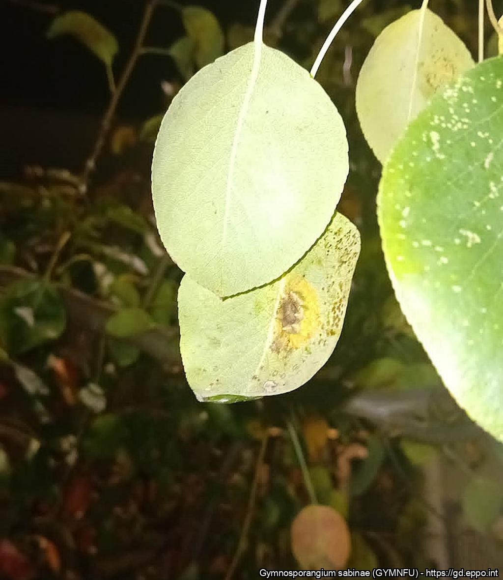
Spermogonia of Gymnosporangium sabinae on the upper leaf surface of pear.
Courtesy: Yulia Holiachuk, Lviv National Environmental University, Ukraine
Aecia of Gymnosporangium sabinae on the lower leaf surface of pear
Courtesy: Yulia Holiachuk, Lviv National Environmental University, Ukraine
Spermogonia of Gymnosporangium sabinae on the upper leaf surface of pear.
Courtesy: Yulia Holiachuk, Lviv National Environmental University, Ukraine
Aecia of Gymnosporangium sabinae on the lower leaf surface of pear.
Courtesy: Yulia Holiachuk, Lviv National Environmental University, Ukraine