Cryphonectria parasitica(ENDOPA)
Photos
For publication in journals, books or magazines, permission should be obtained from the original photographers with a copy to EPPO.
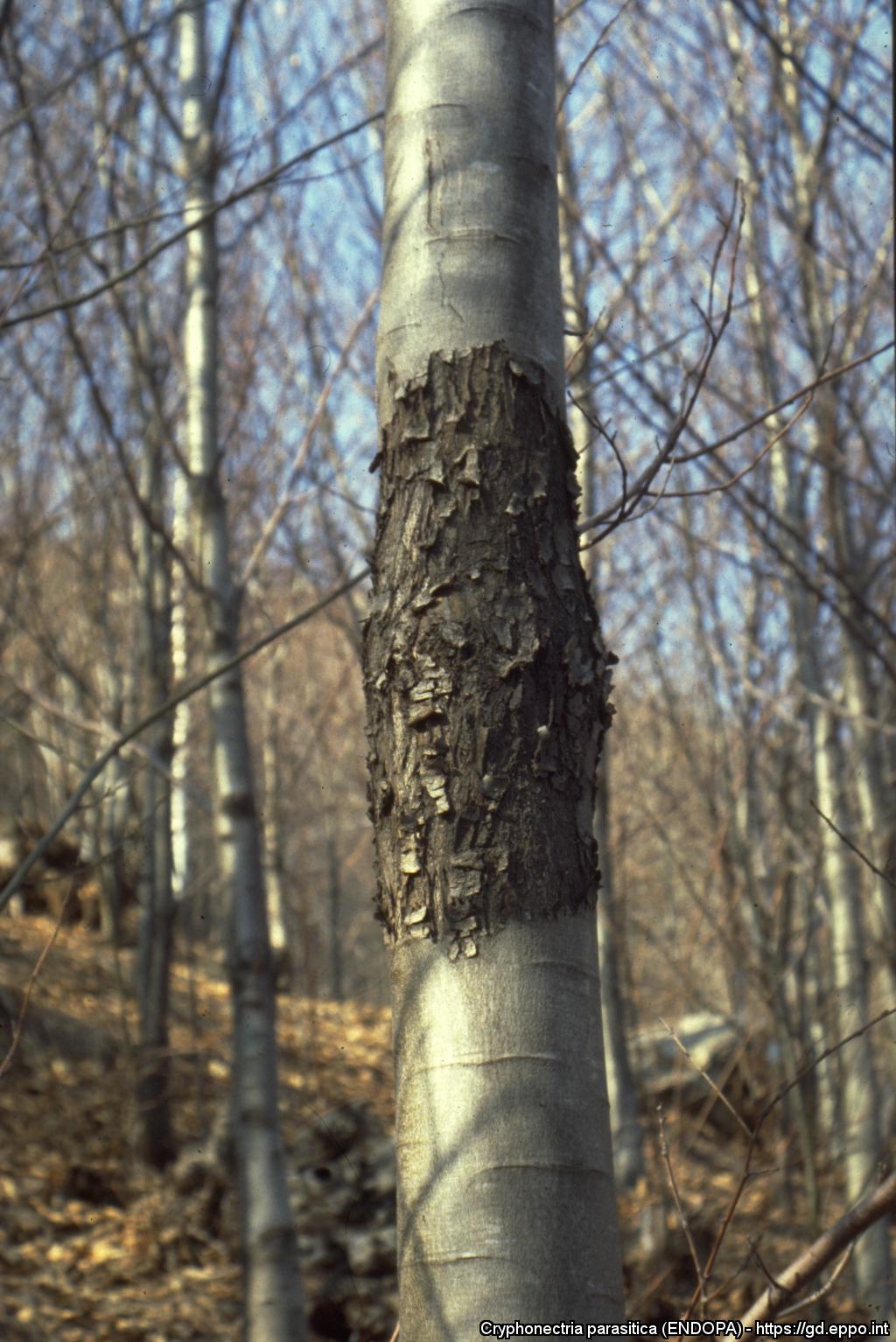
Passive (healed) chestnut blight canker associated with hypovirulence of C. parasitica
Courtesy: Daniel Rigling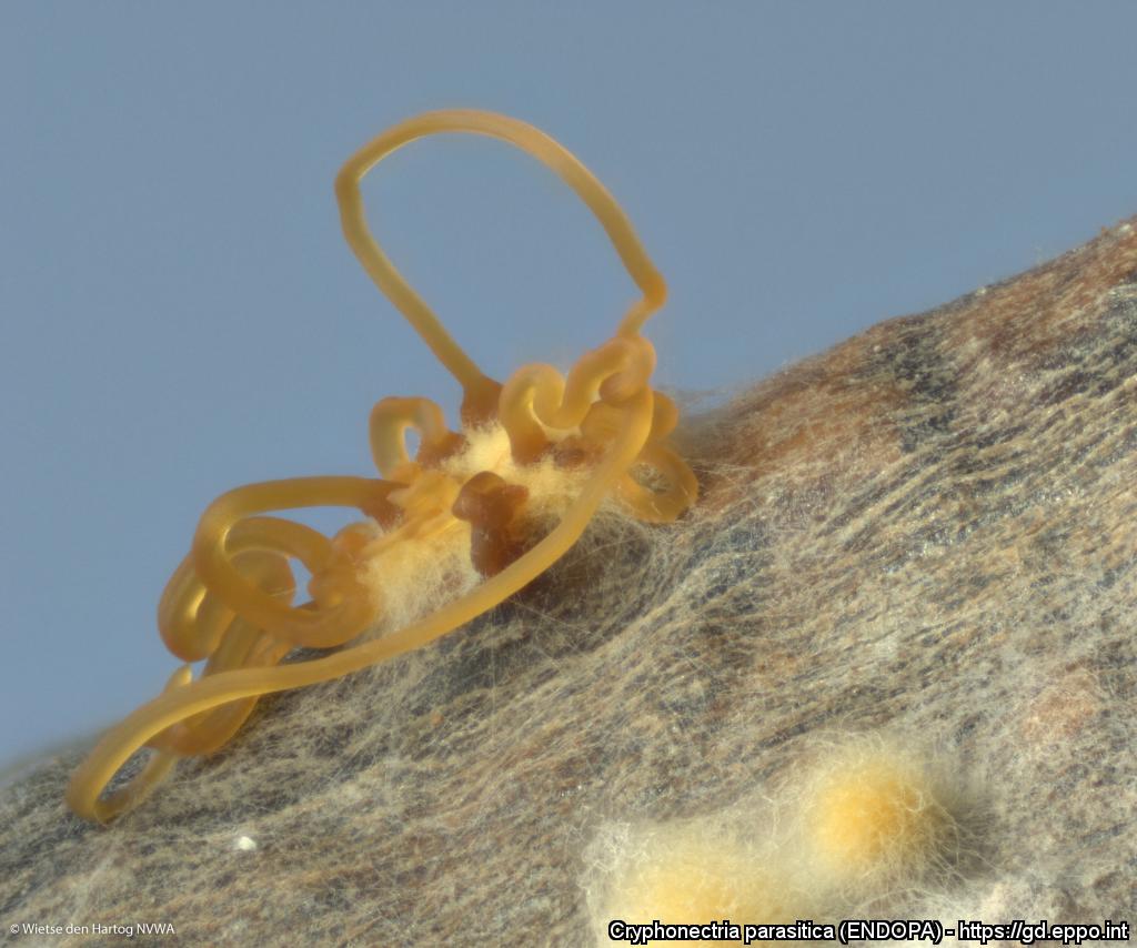
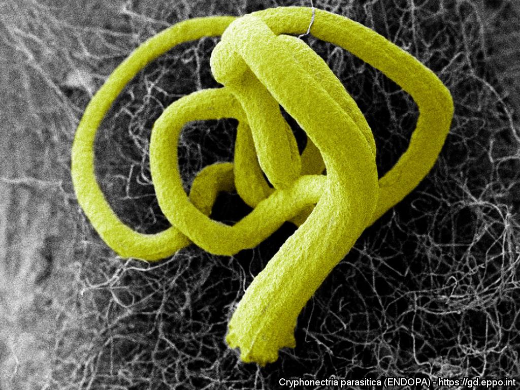
Scanning electron micrograph showing pycnidial ooze of Cryphonectria parasitica
Courtesy: Paul Beales


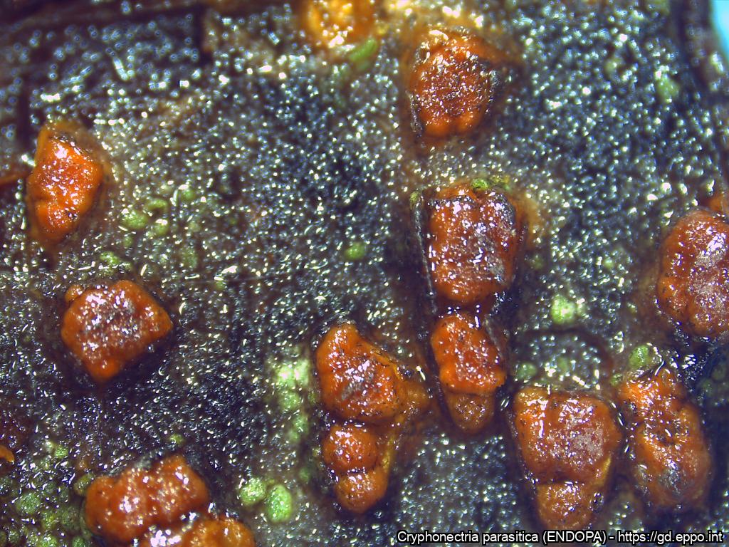
Mature fruiting bodies
Courtesy: Souhila Aouali, Forest Protection Division, Forest Research Institute, Algeria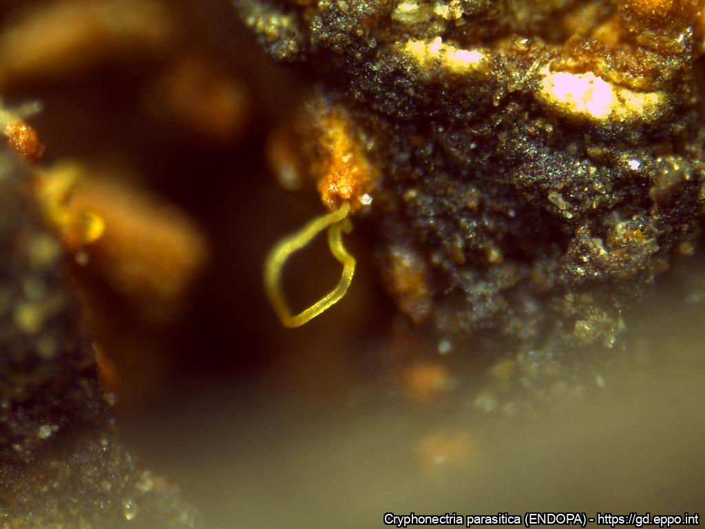
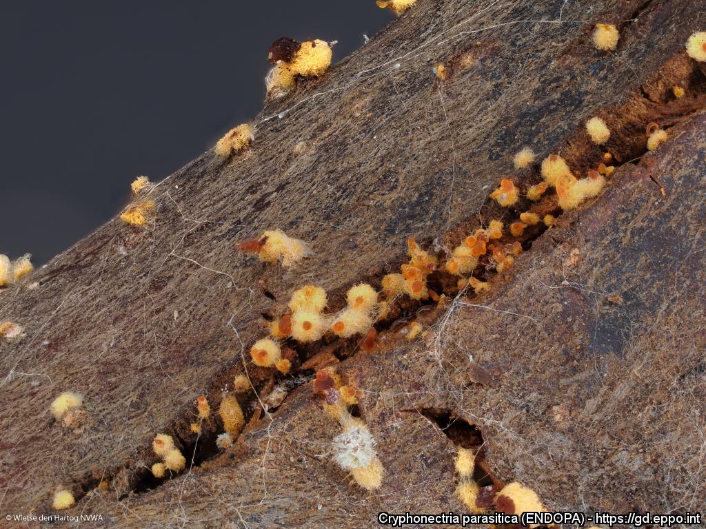
Oozing conidiomata
Courtesy: Souhila Aouali, Forest Protection Division, Forest Research Institute, Algeria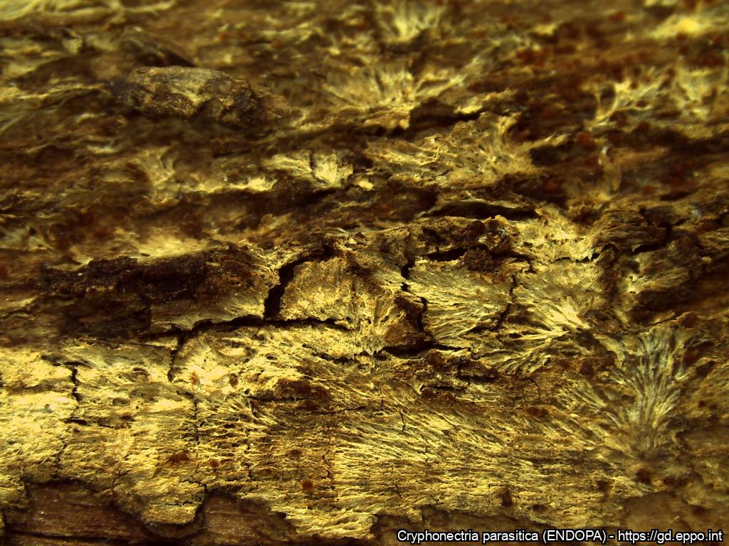
Mycelial fans
Courtesy: Souhila Aouali, Forest Protection Division, Forest Research Institute, Algeria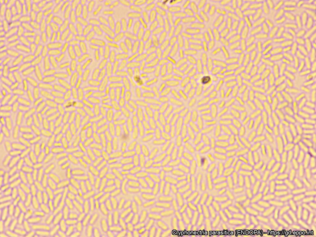
Conidia
Courtesy: Souhila Aouali, Forest Protection Division, Forest Research Institute, Algeria
Cross section of conidiomata
Courtesy: Souhila Aouali, Forest Protection Division, Forest Research Institute, Algeria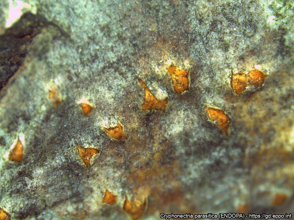
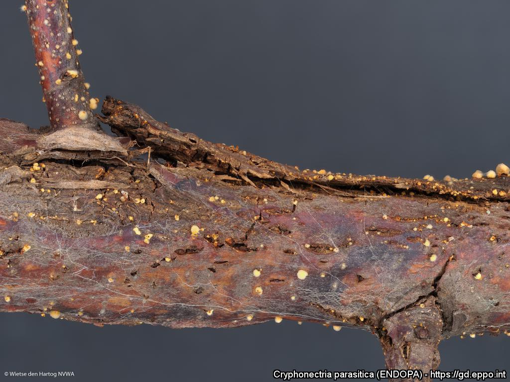
Fruiting bodies of C. parasitica emerging from under the bark
Courtesy: Souhila Aouali, Forest Protection Division, Forest Research Institute, Algeria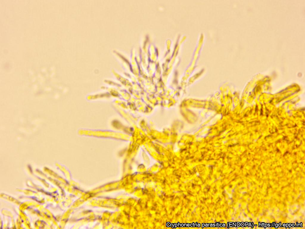
Conidiogenous cells
Courtesy: Souhila Aouali, Forest Protection Division, Forest Research Institute, Algeria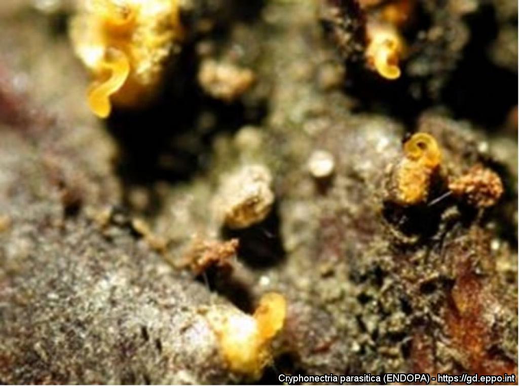
Pycnidial stromata in the bark, conidial tendrils are excreted
Courtesy: LNPV, Nancy (FR)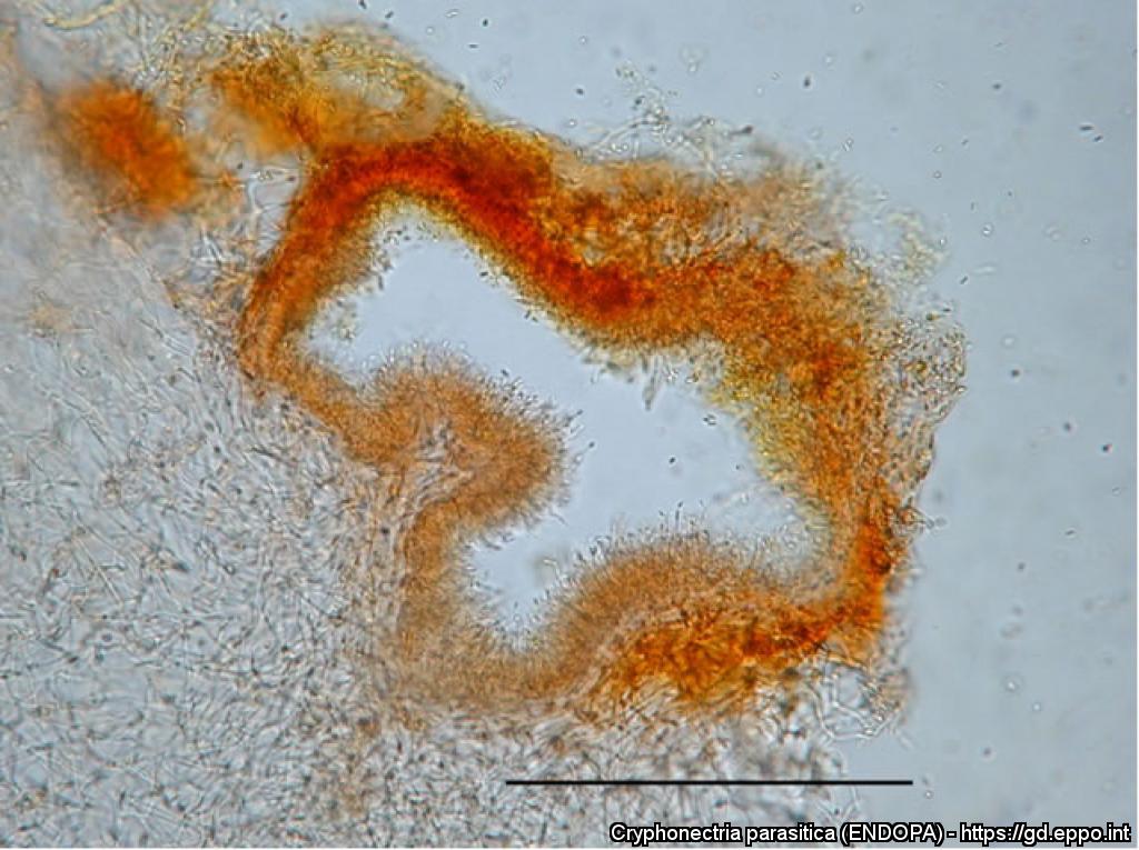
Pycnidium from culture, inner wall is covered with conidiogenous cells (bar = 50 ìm)
Courtesy: SFI, Ljubljana (SI)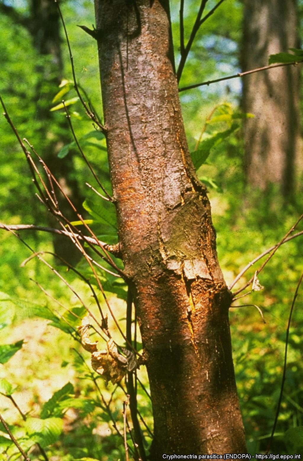
Characteristic mycelial fan of C. parasitica in the inner bark of a chestnut tree
Courtesy: Ministry of Agriculture (HU)
Long, orange-yellow tendrils of C. parasitica pycnidiospores exuding from pycnidia on chestnut tree bark
Courtesy: Ministry of Agriculture (HU)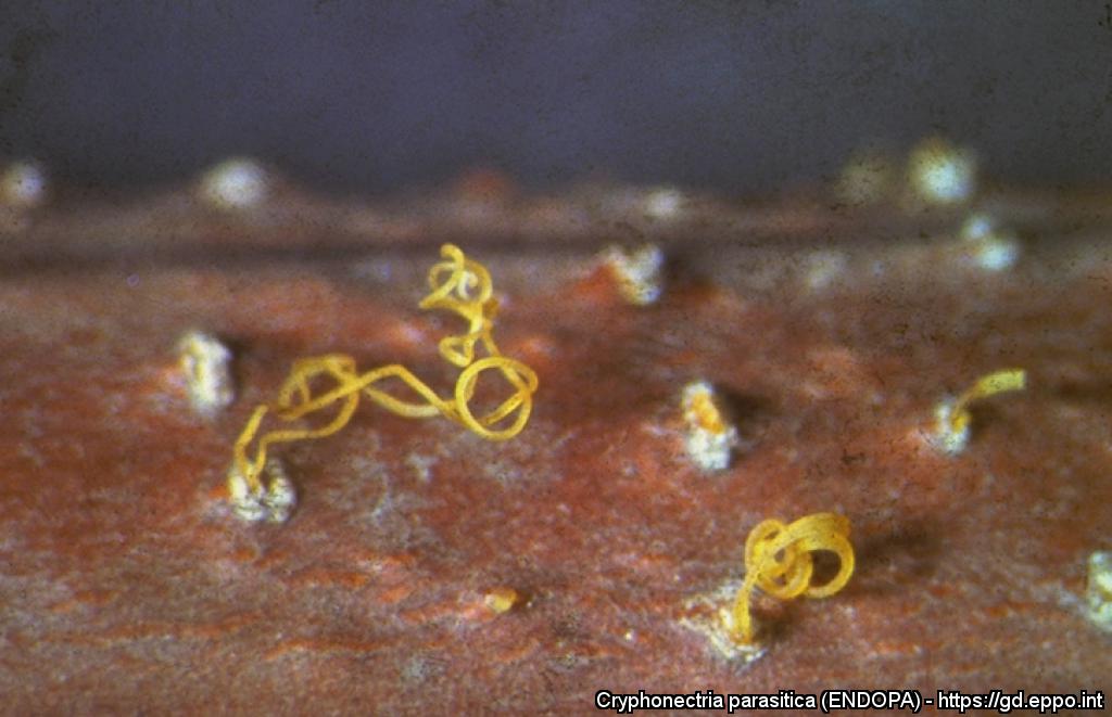
Orange-yellow pycnidia of C. parasitica on chestnut tree bark
Courtesy: Ministry of Agriculture (HU)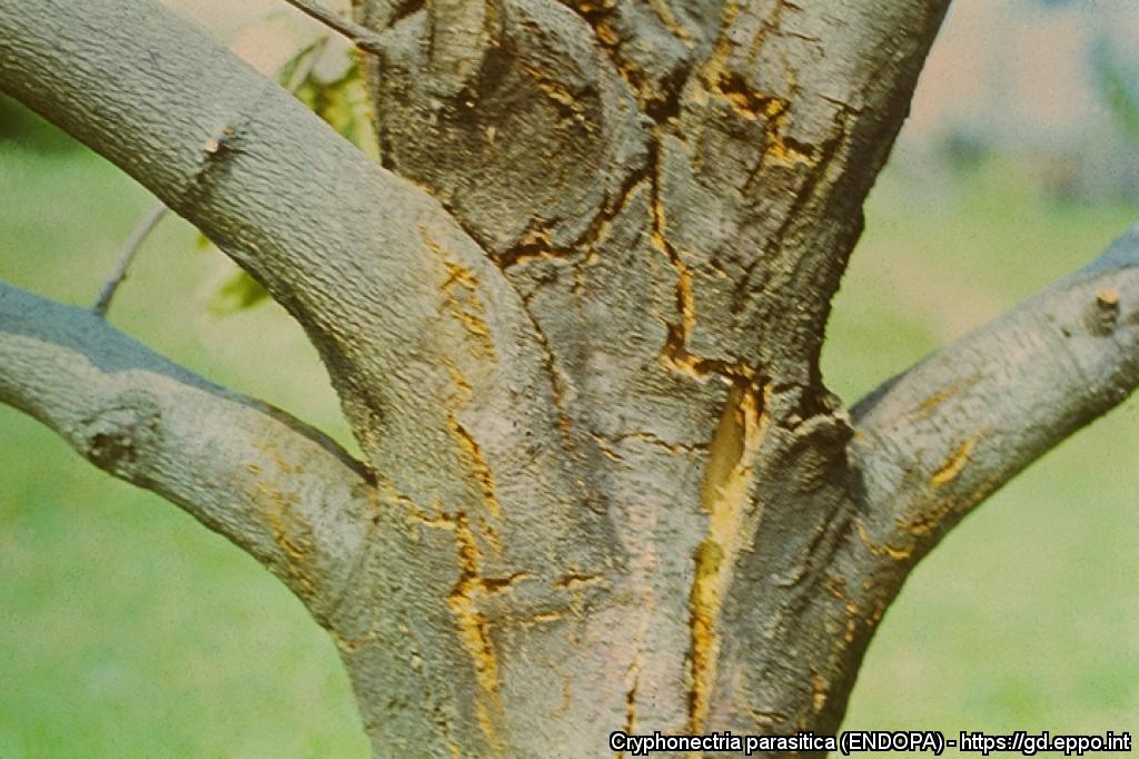
Swelling and bark cracking on 13-year-old chestnut tree
Courtesy: Ministry of Agriculture (HU)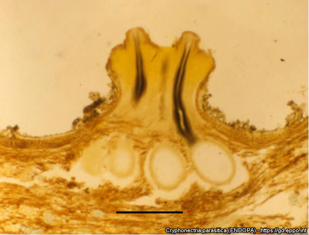
Perithecia in a stroma, necks and ostioles in papillate protuberances of the stroma (bar = 500 ìm)
Courtesy: SFI, Ljubljana (SI)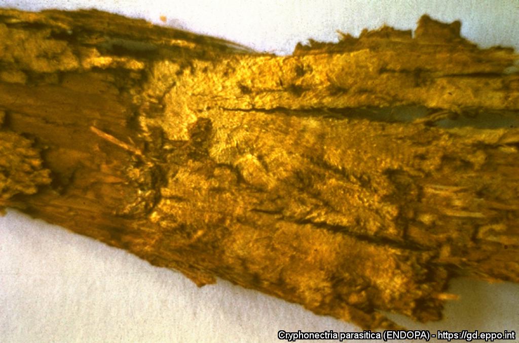
Sunken area on bark of a young chestnut tree. Note adventitious shoots arising below the dead patch
Courtesy: Ministry of Agriculture (HU)
Canker with orange discoloration
Courtesy: Helena Bragança, INIAV (PT)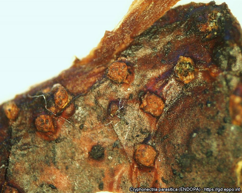
Perithecia
Courtesy: Helena Bragança, INIAV (PT)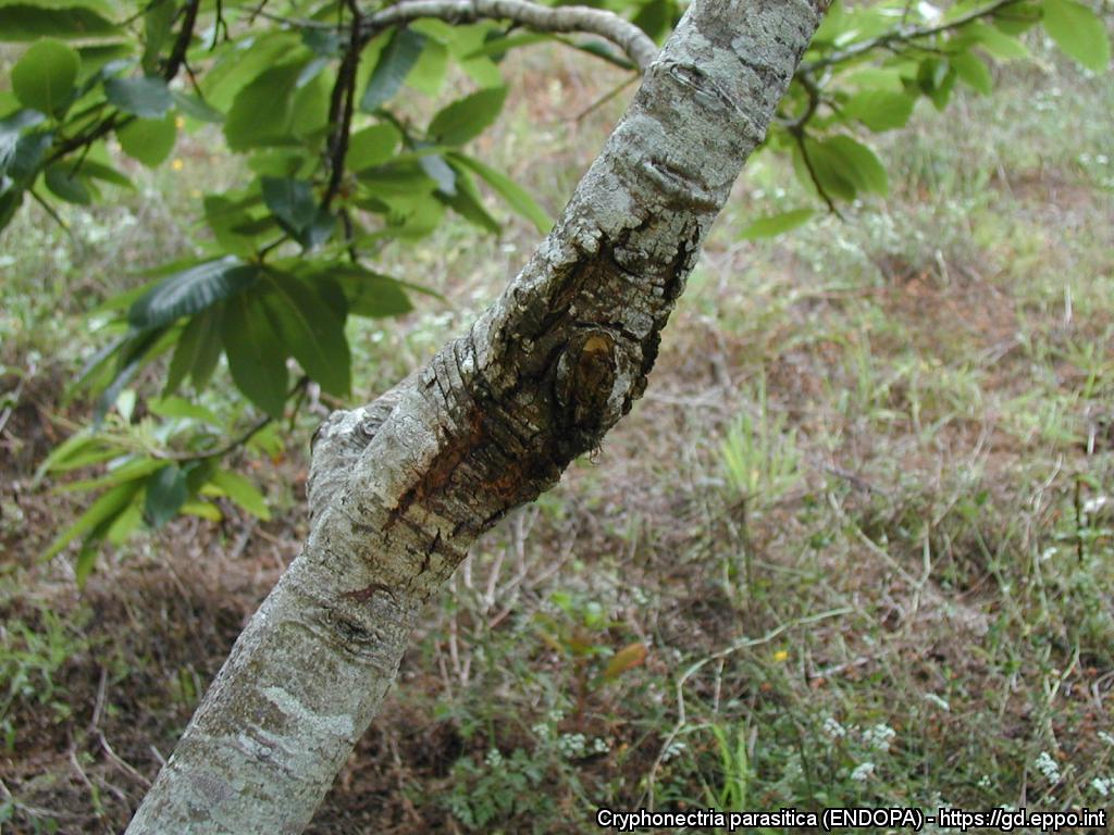
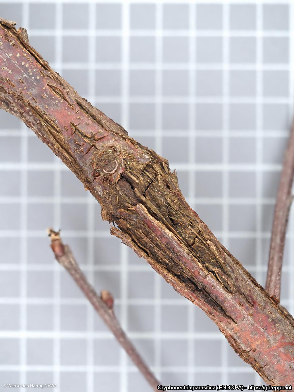
Canker
Courtesy: Helena Bragança, INIAV (PT)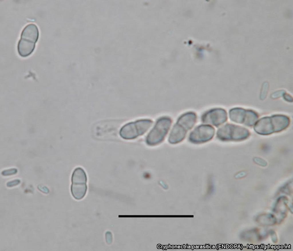
Ascus, ascospores and conidia (bar = 20 ìm)
Courtesy: SFI, Ljubljana (SI)
Cultures of C. parasitica isolates on PDA (1 – virulent, 2 – hypovirulent, 3 and 4 – intermediate virulence)
Courtesy: SFI, Ljubljana (SI)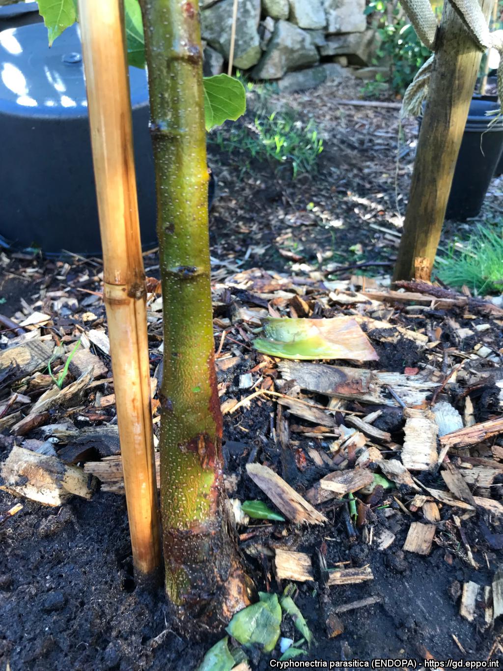
Canker caused Cryphonectria parasitica
Courtesy: Carina Alaby Dieden, Swedish NPPO
Canker caused Cryphonectria parasitica
Courtesy: Carina Alaby Dieden, Swedish NPPO