Benyvirus necrobetae(BNYVV0)
Photos
For publication in journals, books or magazines, permission should be obtained from the original photographers with a copy to EPPO.
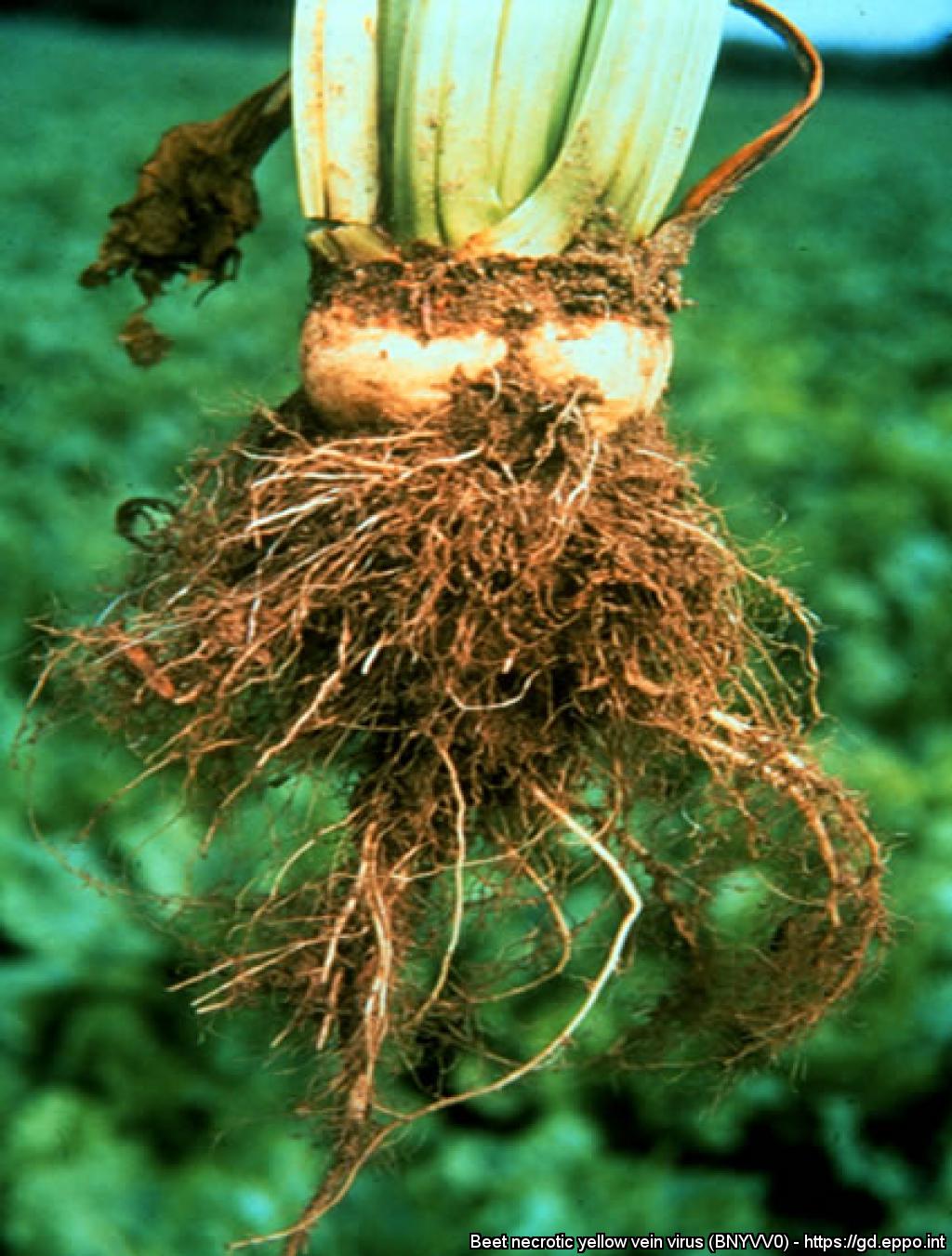
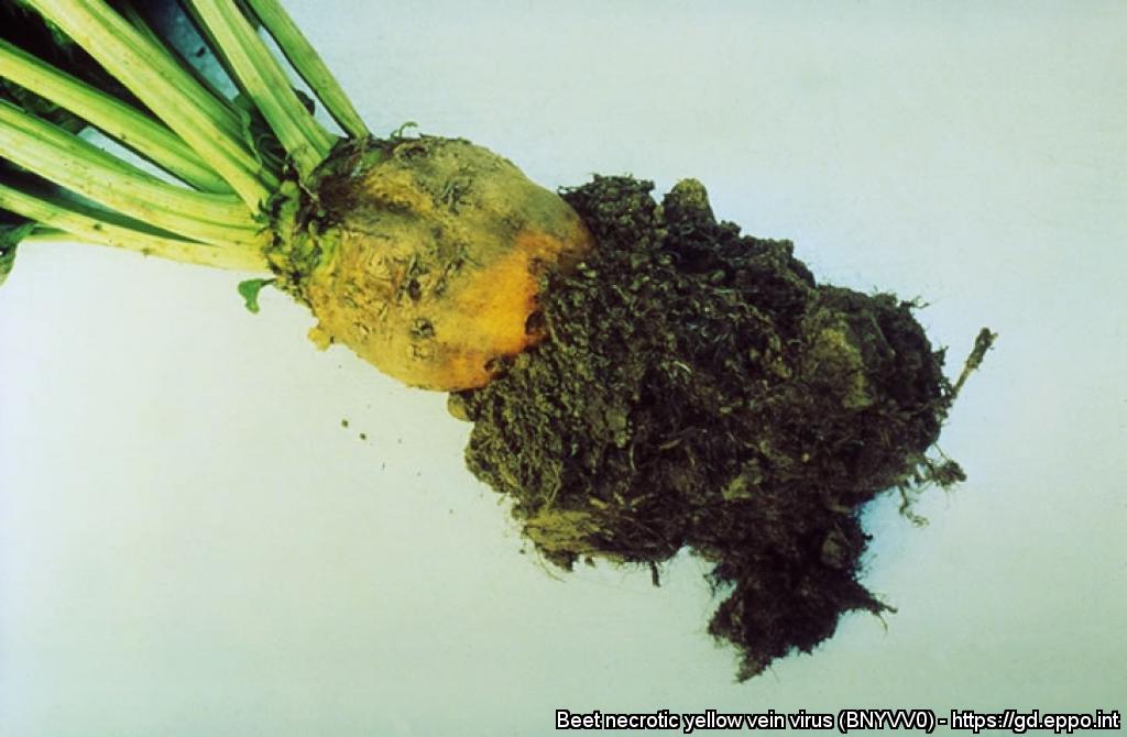
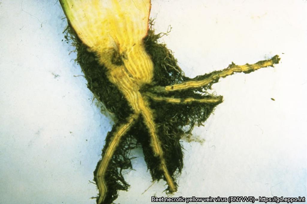
Typical external root symptoms of rhizomania showing the reduced size of the beet and root proliferation (bearding).
Courtesy: Central Science Laboratory, York (GB) - British Crown.
Rhizomania Pocket Diagnostic lateral flow test kit. Top: lateral flow device- negative Bottom: lateral flow device- positive.
Courtesy: Central Science Laboratory, York (GB) - British Crown.
Foliar symptoms of rhizomania: pale green leaves, upright foliage, narrowed leaf laminae.
Courtesy: Central Science Laboratory, York (GB) - British Crown.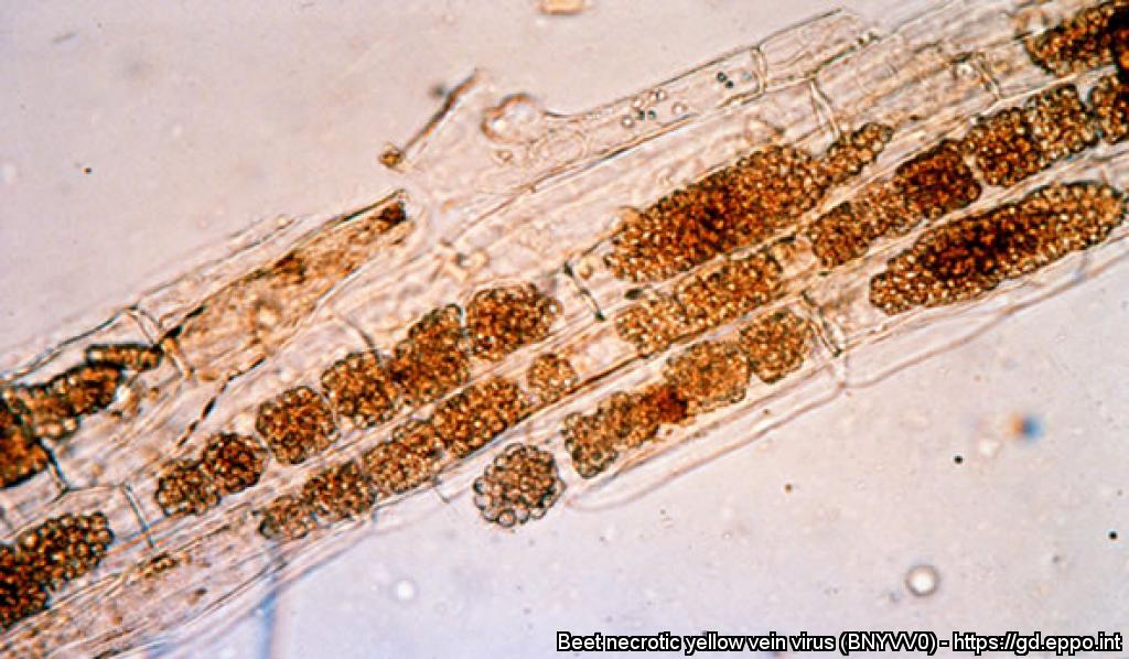
Microscope slide of Polymyxa betae cystosori.
Courtesy: Central Science Laboratory, York (GB) - British Crown.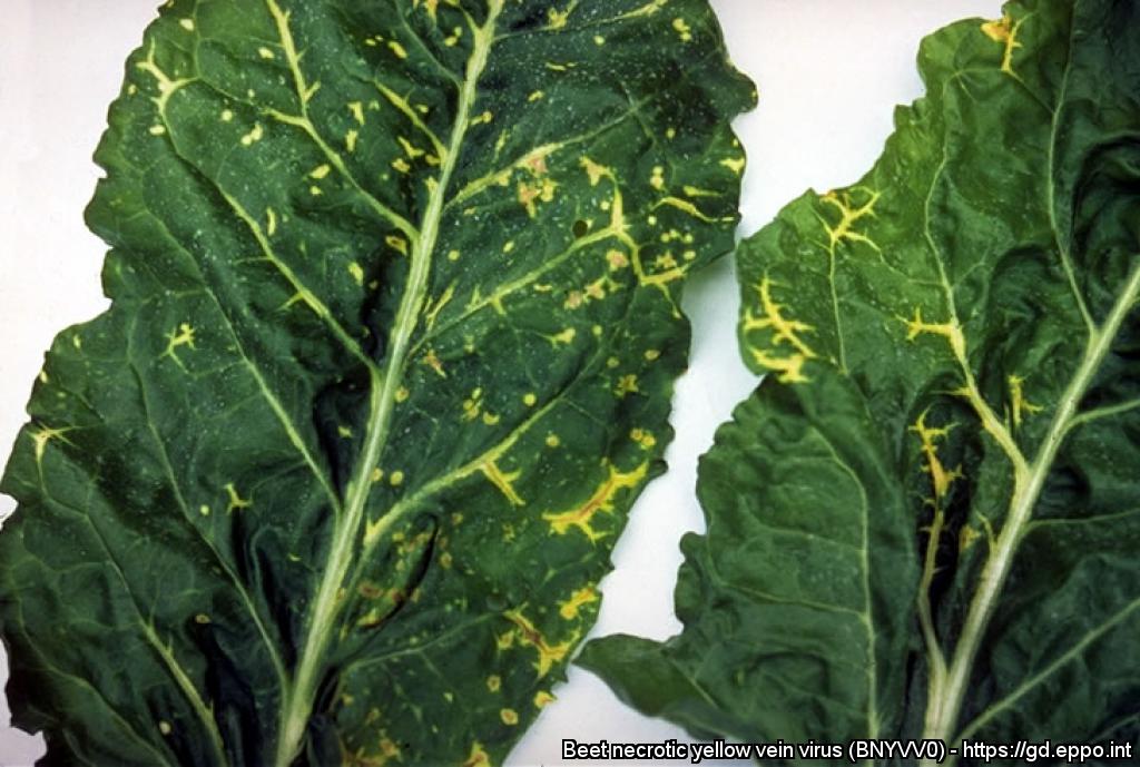
Systemic symptoms on sugar beet leaves.
Courtesy: Institut Technique de la Betterave, France.
Foliar symptoms of rhizomania: yellow veining following the midrib of the leaf. Central Science Laboratory, York (GB) - British Crown
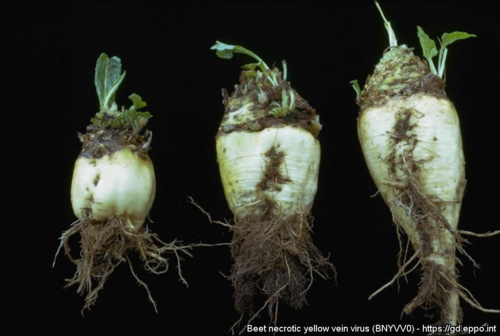
Abnormal root development (rhizomania) in beet.
Characteristic rootlet proliferation, or rhizomania, in sugar beet.
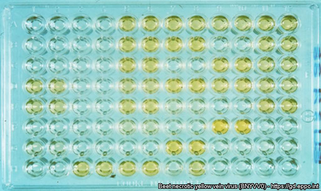
ELISA diagnostic test: yellow wells, when using alkaline phosphatase substrate, indicate a positive result - BNYVV is present.
Courtesy: Central Science Laboratory, York (GB) - British Crown.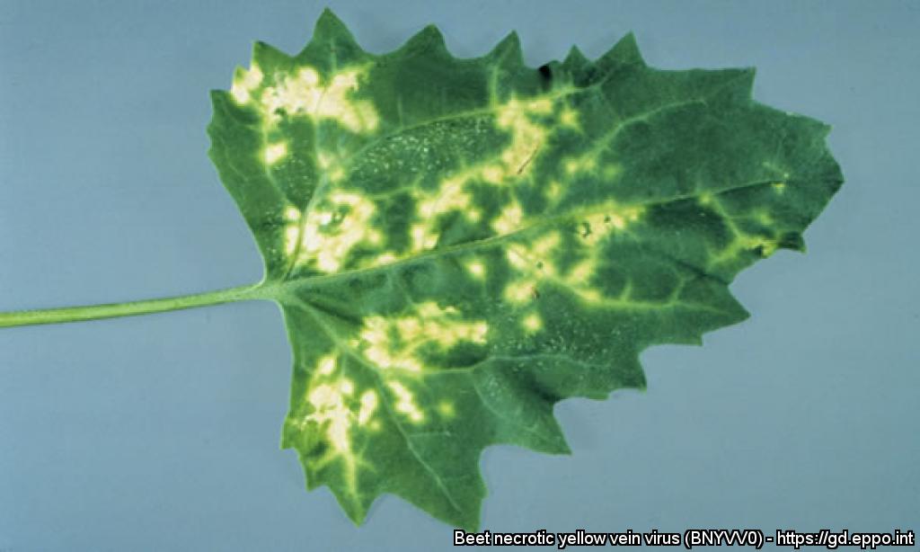
Chlorotic lesions of BNYVV in Chenopodium quinoa.
Courtesy: Central Science Laboratory, York (GB) - British Crown.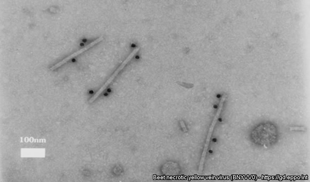
An Electron micrograph of immunogold - labelling of BNYVV virus particles.
Courtesy: Central Science Laboratory, York (GB) - British Crown.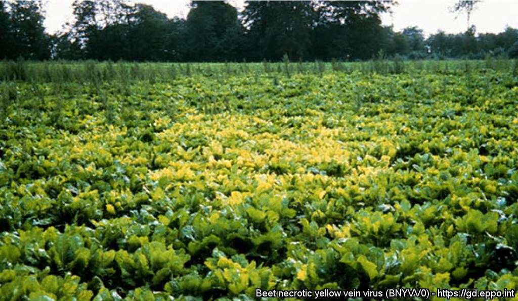
Typical symptoms of rhizomania in the field: a distinct yellow patch of infected sugar beet.
Courtesy: Central Science Laboratory, York (GB) - British Crown.
Electron micrograph of rod-shaped virions of BNYVV, using the IEM method.
Longitudinal section of sugar beet root, showing browning of the vascular tissues.
Courtesy: Institut Technique de la Betterave, France.